
The aim of this study is to define the predictors of chronic carditis in patients with acute rheumatic carditis (ARC).
MethodsPatients diagnosed with ARC between May 2010 and May 2011 were included in the study. Echocardiography, electrocardiography, lymphocyte subset analysis, acute phase reactants, plasma albumin levels, and antistreptolysin-O (ASO) tests were performed at initial presentation. The echocardiographic assessments were repeated at the sixth month of follow-up. The patients were divided into two groups according to persistence of valvular pathology at 6th month as Group 1 and Group 2, and all clinical and laboratory parameters at admission were compared between two groups of valvular involvement.
ResultsDuring the one-year study period, 22 patients had valvular disease. Seventeen (77.2%) patients showed regression in valvular pathology. An initial mild regurgitation disappeared in eight patients (36.3%). Among seven (31.8%) patients with moderate regurgitation initially, the regurgitation disappeared in three, and four patients improved to mild regurgitation. Two patients with a severe regurgitation initially improved to moderate regurgitation (9.1%). In five (22.8%) patients, the grade of regurgitation [moderate regurgitation in one (4.6%), and severe regurgitation in 4 (18.2%)] remained unchanged. The albumin level was significantly lower at diagnosis in Group 2 (2.6±0.48g/dL). Lymphocyte subset analysis showed a significant decrease in the CD8 percentage and a significant increase in CD19 percentage at diagnosis in Group 2 compared to Group 1.
ConclusionThe blood albumin level and the percentage of CD8 and CD19 (+) lymphocytes at diagnosis may help to predict chronic valvular disease risk in patients with acute rheumatic carditis.
O objetivo deste estudo é definir os preditores da cardite crônica em pacientes com cardite reumática aguda (CRA).
MétodosOs pacientes diagnosticados com CRA entre maio de 2010 e maio de 2011 foram incluídos no estudo. Foram realizados os testes de ecocardiografia, eletrocardiograma, uma análise do subgrupo de linfócitos, provas de fase aguda, níveis de albumina plasmática, antiestreptolisina-O (ASO) na manifestação inicial. As avaliações ecocardiográficas foram repetidas no 6° mês de acompanhamento. Os pacientes foram divididos em dois grupos de acordo com a persistência da patologia valvular no 6° mês como Grupo 1 e Grupo 2 e todos os parâmetros clínicos e laboratoriais na internação foram comparados entre dois grupos de comprometimento valvular.
ResultadosDurante o período do estudo de um ano, 22 pacientes apresentaram doença valvular. 17 (77,2%) pacientes apresentaram regressão da patologia valvular. Houve desaparecimento de regurgitação moderada inicial em 8 pacientes (36,3%). Entre 7 (31,8%) pacientes com regurgitação moderada inicialmente, a regurgitação desapareceu em 3 e 4 pacientes apresentaram melhora para regurgitação leve. 2 pacientes com regurgitação grave inicialmente apresentaram melhora para regurgitação moderada (9,1%). Em 5 (22,8%) pacientes, o grau de regurgitação [regurgitação moderada em 1 (4,6%) e regurgitação grave em 4 (18,2)] continuou inalterado. O nível de albumina foi significativamente menor no diagnóstico no Grupo 2 (2,6±0,48 gr/dL). A análise do subgrupo de linfócitos mostrou uma redução significativa no percentual de CD8 e um aumento significativo no percentual de CD19 no Grupo 2 em comparação ao Grupo 1.
ConclusãoO nível de albumina no sangue e o percentual de linfócitos CD8 e CD19 (+) no diagnóstico podem ajudar a prever risco de doença valvular crônica em pacientes com cardite reumática aguda.
The latent period of ARF (acute rheumatic fever) following streptococcal infections, infiltration of T-cells and macrophages in heart valves, and B-cell infiltration in Aschoff nodules indicate that the immune system is involved in disease pathogenesis.1 Studies investigating the alterations in cellular immune response during ARF have shown decreased CD3-CD8 (+) lymphocytes and increased CD4/CD8 lymphocyte ratio.2 CD8 levels were reported to decrease in patients with rheumatic heart disease compared to patients with ARF or healthy subjects, indicating an immune process in the course of this disease, and CD8 T-cells have been shown to play an important immunoregulatory role in the pathogenesis.3
B-cells perform roles such as antigen presentation, cytokine production, and the regulation of lymphoid organogenesis, effector T-cell differentiation, and dendritic cell function.4 Accordingly, critical roles of B-cells have been demonstrated in several immune-mediated diseases. Because the fate and function of B-cells are tightly regulated by signal transduction through B-cell receptors and functionally interrelated cell surface receptors, such as CD19, CD21, CD22, CD40 can be a potential strategy for regulating these disorders.5,6 Among them, CD19 is generally considered as a positive B cell response regulator.7
The plasma albumin level may decrease in inflammatory conditions. Hypoalbuminemia lowers plasma oncotic pressure; it may contribute to pulmonary edema in the presence of elevated left atrial pressure and contribute to the progression of heart failure, and it is commonly seen in patients with heart failure.8 Several studies have demonstrated a low survival rate and a high rate of cardiovascular death in patients with hypoalbuminemia (<3.4g/dL),9 and one study has emphasized that serum albumin <2.5g/dL as the most independent predictor of in-hospital mortality.10 However, the albumin-ARF relationship has never been investigated for chronic valvular disease in children.
The present study aimed to define predictors of chronic carditis through evaluation of electrocardiography, echocardiography, lymphocyte subset analysis, and laboratory parameters including albumin, ESR, CBC, and ASO in patients diagnosed with acute rheumatic carditis prior to receiving treatment with steroids.
Materials and methodsTwenty-two patients admitted to the pediatric cardiology unit with a diagnosis of acute rheumatic carditis between May 2010 and May 2011 were included in this retrospective study. Complete blood count, plasma albumin level, C-reactive protein (CRP), erythrocyte sedimentation rate (ESR), antistreptolysin O (ASO), lymphocyte subset analysis, echocardiography, and electrocardiography were performed before treatment in eligible patients. The study was approved by the institutional review board.
All patients with presumed diagnosis of ARF were evaluated by echocardiography regardless of cardiac auscultation findings. The diagnosis of ARF was based on the modified Jones criteria (2 major or 1 major+2 minor and previous Group A beta hemolytic streptococcus (GABHS) predictors).11 Valvular regurgitation was considered to be pathologic when a color jet length of at least 1cm was found, seen in at least two separate planes with a peak velocity greater than 2.5m/s in echocardiographic examination. Patients who were diagnosed with ARC for the first time were included into study. Patients with previous diagnosis of ARF and/or ARC were not included. Accordingly, 2mg/kg/d (max. 60mg) prednisone was initiated for all patients. After two to four weeks of prednisone, the dosage was tapered over a two-week period. Acetylsalicylic acid was added to the treatment during steroid taper at a dose of 90–100mg/kg/d (max. 3.5g) and was discontinued after taper, with an overall duration of four weeks.
An injection of 1,200,000U benzathine penicillin G for patients who weigh more than 27kg and 600,000U intramuscular for patients who weigh 27kg or less every three weeks is the recommended regimen for secondary prevention.
At the time of diagnosis, eight patients had mild regurgitation. A moderate and severe regurgitation was present in eight and six patients, respectively. No patients experienced a recurrent episode of carditis during follow-up period. After steroid therapy, acute phase reactants remained negative throughout the study. The patients were divided into two groups, as those with (Group 1) and without (Group 2) regression in valvular regurgitation according to the echocardiographic assessment at post-treatment month six, and the data of these groups at the initial diagnosis were compared. Access to the hospital database was granted by the hospital administration.
EchocardiographyTransthoracic two-dimensional and Doppler echocardiographic evaluations of the subjects were performed with the GE Vivid 3 device (GE Healthcare–Milwaukee, WI, United States) using 3MHz and 7MHz transducers for cardiac assessment. M-mode echocardiography measurements were obtained from the level of the posterior mitral valve according to the recommendations of the American Society of Echocardiography. The severity of valvular regurgitation was assessed by color-Doppler echocardiography using the width and extent of the jet flow. The criteria for valvular regurgitation using orthogonal planes were a color jet observed in at least two separate planes, a color jet length of at least 1cm, and a color jet mosaic with a peak velocity greater than 2.5m/s. The severity of mitral regurgitation and aortic regurgitation were graded qualitatively from 1 to 4 depending on the observed regurgitant jet flow in the left atrium or the left ventricle by color-Doppler echocardiography. The mitral regurgitation and aortic regurgitation detected by color-Doppler was considered grade 1 if the jet length was 1.5cm; grade 2 if 1.5–2.9cm; grade 3 if 3–4.4cm; and grade 4 if 4.5cm.12 Grade 1 was considered as mild, grade 2 as moderate, and grades 3 and 4 as severe regurgitation.13,14
Electrocardiography12-lead surface ECG recordings were obtained from all patients. The QT interval was measured from the beginning of the Q wave to the end of the T wave, defined as the point at which the T wave converted to the isoelectric line. When a U wave was present, the end of the T wave was defined as the lowest point between the T and U waves. QT dispersion was defined as the difference between the maximal and minimal QT intervals on 12-channel standard ECG. The QT intervals on ECGs were corrected using Bazett's formula (QTc=QT/√R−R) and were expressed as QTc.15
Flow cytometryLymphocyte subgroups were measured with the indirect immunofluorescence method, using monoclonal antibodies. The method involves the steps of staining with monoclonal antibodies, incubation, removal of erythrocytes, and material fixation with paraformaldehyde. Monoclonal antibodies are prepared as a panel and contain fluorochrome. There are many fluorochromes with different spectral characteristics; the suspended cells or particles are passed through a chamber illuminated by a laser light, and the signals given by the cells as they pass by the light are collected and analyzed. The signal source may be the physical characteristics of the cell, such as the magnitude and granularity, as well as various fluorochromes bound to the cell. Thereby, data can be collected regarding several properties of the cell or particle, such as immunophenotype, DNA content, enzyme activities, cellular membrane potential, and viability. The presence of an entity (antigen) in the cell can be immunologically demonstrated by immunohistochemistry methods through a stain, an enzyme attached to a protein produced in response to such entity (antibody) or by immunofluorescent microscopy through a fluorescent substance. The cellular surface antigens are defined using the cluster of differentiation (CD) terminology.16 Blood samples were tested in the hematology laboratory. Flow cytometry analysis was performed with blood collected in ethylenediaminetetraaceticacid (2mg/mL) tubes using a Beckman Coulter FC 500 device. CD3, CD4, CD8, and CD19 percentages were calculated.
Laboratory testsKnown as inflammation markers and identified as independent risk factors for heart failure in rheumatic diseases, the following were studied: C-reactive protein (CRP) and erythrocyte sedimentation rate (ESR);17 plasma albumin, a negative inflammation indicator associated with cardiac mortality;10 antistreptolysin (ASO) level, as an indicator of GABHS infection. The calorimetric bromocresol green method was used to assess albumin levels. C-reactive protein (CRP) concentrations and ASO levels were studied nephelometrically using a Behring kit. Sedimentation was measured using the Westergren method.
Statistical assessmentStatistical analyses were performed using the software SPSS for Windows v. 17.0 (SPSS; Chicago, IL, USA). Normal distribution of the variables was assessed with the Kolmogorov–Smirnov test. The t-test was used to investigate the significance of differences between the control and the patient groups for the series with regular distribution, and the Mann–Whitney U test was used for the series with irregular distribution. p-values below 0.05 were considered statistically significant.
ResultsTwenty-two patients (12 male, 10 female, mean age of 12.23±2.1years) with diagnosis of acute rheumatic carditis were included into the study. None of the patients with carditis had Sydenham chorea, erythema marginatum, or subcutaneous nodules. Detailed clinical and laboratory data are presented in Table 1. Echocardiographic and electrocardiographic assessment revealed mean fractional shortening, mean heart rate, QTc dispersion, and PR distance as 42.9%±4.5%, 91.10±20.27/min, 42.7±17.5ms, and 0.15±0.04s, respectively (Table 1). Electrocardiographic characteristics demonstrated no difference in terms of PR interval, mean heart rate, and QTc dispersions between Group 1 and Group 2 (p>0.05; Table 2).
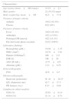
Demographic, clinical characteristics, and laboratory parameters of the patients at the time of diagnosis.
| Characteristics | |
|---|---|
| Age (years), mean±SD (range) | 12.23±2.1 |
| Male gender | 22 (54.5% male) |
| Body weight (kg), mean±SD | 42.4±17.9 |
| Presence of major criteria | |
| Arthritis | 18/22 (81.8%) |
| Chorea | 0/22 (0%) |
| Presence of minor criteria | |
| Fever | 10/22 (45.4%) |
| Arthralgia | 3/22 (13.6%) |
| Prolonged PR interval | 4/22 (18.1%) |
| Elevated acute phase reactants | 22/22 (100%) |
| Laboratory findings | |
| Hemoglobin (g/dL) | 10.49±1.35 |
| WBC (/mm3) | 10.79±3.91 |
| Platelet (1000/mm3) | 409±115 |
| ESR (h) | 106±26 |
| ASO (IU/mL) | 1175±425 |
| Albumin (g/dL) | 3.21±0.55 |
| Echocardiography | |
| FS% | 42.9±4.5 |
| Electrocardiography | |
| Heart rate (pulse/min) | 91.10±20.27 |
| QTc dispersion (ms) | 42.7±17.5 |
| PR distance (sec) | 0.15±0.04 |
| Lymphocyte subset analysis | |
| CD8 (%) | 25.65±6.14 |
| CD19 (%) | 21.11±8.72 |
ASO, anti-streptolysine O; ESR, erythrocyte sedimentation rate; FS, fractional shortening; WS, white blood cell count.
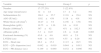
Comparison of electrocardiography–echocardiography, and laboratory findings of the groups with and without regression in carditis.
| Variable | Group 1 | Group 2 | p |
|---|---|---|---|
| Number | 17 (77.2%) | 5 (22.8%) | |
| Age range (mean/years) | 12±1.8 | 13±3.2 | NS |
| Sedimentation (h) | 104.1±25.7 | 113.6±29.4 | NS |
| ASO (IU/mL) | 1192±438 | 1118±418 | NS |
| White blood cell (/mm3) | 10.27±3.9 | 12.56±3.58 | NS |
| Hemoglobin (g/dL) | 10.75±1.36 | 9.6±0.91 | NS |
| Platelet (1000/mm3) | 397±92 | 451±182.3 | NS |
| Albumin (g/dL) | 3.3±0.45 | 2.6±0.48 | 0.003 |
| Fractional shortening (%) | 43.6±4.6 | 40.0±2.9 | NS |
| LVEDd (cm) | 4.66±0.61 | 4.55±0.20 | NS |
| ECG – Heart rate | 86.2±15.2 | 111.5±28.5 | NS |
| ECG – QTc dispersion (sec) | 0.042±0.019 | 0.044±0.011 | NS |
| ECG – PR distance (sec) | 0.149±0.040 | 0.152±0.046 | NS |
ASO, anti-streptolysine O; ECG, electrocardiography; LVEDd, left ventricular end diastolic diameter; NS, non-significant.
Routine blood tests demonstrated no association in hemoglobin, sedimentation, and ASO antibody levels between Group 1 and Group 2, whereas the blood albumin level at diagnosis was found significantly lower in Group 2 (2.6±0.48; Table 2).
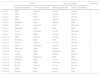
The echocardiographic assessment revealed combined valve involvement in 68.2% of the patients, isolated mitral regurgitation in 22.7%, and isolated aortic regurgitation in 9.1%. Seventeen (77.2%) patients showed regression in valvular pathology, whereas in five (22.8%) patients, the grade of regurgitation (moderate regurgitation in 1 (4.6%), and severe regurgitation in 4 (18.2%) remained unchanged (Table 2). At the time of diagnosis, eight patients had mild regurgitation. A moderate and severe regurgitation was present in eight and six patients, respectively. At the sixth month of follow-up, an initial mild regurgitation disappeared in eight patients (36.3%). Among seven (31.8%) patients with moderate regurgitation initially, the regurgitation disappeared in three patients and four patients improved to mild regurgitation. Two patients with severe initial regurgitation improved to moderate regurgitation (9.1%). In five (22.8%) patients, the grade of regurgitation [moderate regurgitation in 1 (4.6%), and severe regurgitation in four (18.2%) remained unchanged. The fate of valvular involvement in all study patients is depicted on Table 3.
Severity of valvular involvement in all patients at the beginning of the study and at the sixth month of follow-up.
| Initial | After six-months | Recovery | |||
|---|---|---|---|---|---|
| Mitral regurgitation | Aortic regurgitation | Mitral regurgitation | Aortic regurgitation | ||
| Case 1 | Mild | Absent | Absent | Absent | + |
| Case 2 | Mild | Mild | Absent | Absent | + |
| Case 3 | Mild | Mild | Absent | Absent | + |
| Case 4 | Mild | Absent | Absent | Absent | + |
| Case 5 | Moderate | Mild | Absent | Mild | + |
| Case 6 | Mild | Moderate | Mild | Moderate | − |
| Case 7 | Moderate | Mild | Mild | Mild | + |
| Case 8 | Severe | Moderate | Severe | Moderate | − |
| Case 9 | Absent | Mild | Absent | Absent | + |
| Case 10 | Mild | Absent | Absent | Absent | + |
| Case 11 | Moderate | Mild | Mild | Absent | + |
| Case 12 | Severe | Absent | Severe | Absent | − |
| Case 13 | Mild | Moderate | Absent | Absent | + |
| Case 14 | Moderate | Mild | Absent | Absent | + |
| Case 15 | Severe | Mild | Moderate | Mild | + |
| Case 16 | Absent | Mild | Absent | Absent | + |
| Case 17 | Moderate | Moderate | Mild | Mild | + |
| Case 18 | Moderate | Mild | Absent | Absent | + |
| Case 19 | Severe | Mild | Moderate | Absent | + |
| Case 20 | Severe | Absent | Severe | Absent | − |
| Case 21 | Mild | Mild | Absent | Mild | + |
| Case 22 | Severe | Mild | Severe | Mild | − |
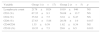
Lymphocyte subset analysis showed lymphopenia in 31.8% (seven) of the patients. Both CD3 and CD4 T-lymphocytes were decreased in 40.9% (nine) of the patients. Furthermore, CD8 T-lymphocytes were also decreased in 13.6% of the patients with decreased levels of both CD3 and CD4. The CD8 percentage at diagnosis significantly decreased in Group 2, whereas there was a significant increase in the CD19 percentage (Table 4).
Comparison of lymphocyte subset analysis of the groups with (group 1) and without (group 2) regression in carditis.
| Variable | Group 1 (n=17) | Group 2 (n=5) | p |
|---|---|---|---|
| Lymphocyte count | 2178±1024 | 1819±946 | NS |
| CD3 (%) | 67.35±9.3 | 58.08±13.7 | NS |
| CD4 (%) | 35.82±7.5 | 32.8±6.47 | NS |
| CD8 (%) | 27.63±5.88 | 20.56±1.9 | 0.017 |
| CD4/CD8 | 1.35±0.39 | 1.62±0.34 | NS |
| CD19 (%) | 19.35±7.5 | 29.6±6.5 | 0.013 |
NS, non-significant.
Various autoantigens with similarity to Group A beta hemolytic streptococcal M proteins have been defined in susceptible subjects: cardiac myosin epitopes, vimentin, laminin, and other intracellular proteins. This similarity causes cross-reactivity leading to autoimmune reactions and may result in myocardial damage. Therefore, several studies have attempted to define these reactions by investigating lymphocyte subgroups.18 Mice immunized with streptococcal M5 protein developed heart valve lesions; CD4 (+) T-cells and CD68 (+) macrophages19 were detected in infiltrations, and predominant infiltration of CD4 T-lymphocytes was found in heart tissue biopsies of patients undergoing valvular surgery due to rheumatic carditis.20 The patients with acute rheumatic fever and acute rheumatic carditis had significantly increased CD4 T-lymphocytes, CD22 B-lymphocytes, and CD4:CD8 ratio and significantly decreased CD8 and CD3 T-lymphocytes compared to the patients with chronic rheumatic carditis, streptococcal pharyngitis, and healthy subjects.2 The decreased CD8 was found to be associated with rheumatic activity in previous studies. In the present study, CD8 T-lymphocytes in peripheral blood at the initial diagnosis was <20.56%±1.9% in the group without recovery in heart valve lesions; this finding was considered an important marker for the group without regression in carditis. The result of the present study might be attributed to the fact that only CD8 T-cells may cause anergy to Group A beta hemolytic streptococcal superantigens and may therefore play an important immunoregulatory role in the pathogenesis of the disease.21 In the present study, the CD4:CD8 ratio increased in the group without recovery in valvular findings compared to the group with recovery; however, the difference was not statistically significant. The percentage of CD19-positive B cells was significantly elevated in ARF patients, suggesting a role of B lymphocytes in inflammation and/or autoimmunity in the pathogenesis of these disorder.22 The present study demonstrated a lower CD8 level in group 2 than group 1 and a higher CD19 level in group 2 than group 1 at diagnosis.
A study by Saito et al. demonstrated that human systemic sclerosis patients overexpress CD19, an important regulatory molecule expressed by B-lymphocytes. B-cells from CD19-deficient mice are hyporesponsive to transmembrane signals, while B-cells overexpressing CD19 are hyperresponsive and generate autoantibodies. Thus, a CD19-dependent signaling pathway in B cells contributes to the development of systemic autoimmunity.23 A study by Iwata et al. was the first to demonstrate that B-cells play a critical role in the wound-healing process. The CD19 expression positively regulates the wound-healing process. Delayed wound healing in CD19-/- mice was associated with decreased infiltration of neutrophils and macrophages.24 In the present study, the CD8 percentage at diagnosis significantly decreased in ARC patients without recovery in valvular involvement, whereas there was a significant increase in the CD19 percentage. Given the role of CD8 and CD19 (+) lymphocytes in autoimmunity and inflammation, it is believed that the alteration of these two subsets of lymphocytes might have a role in pathogenesis and prediction of chronic valvular involvement in patients with ARF.
The blood albumin level may decrease in inflammatory conditions and heart failure. The potential causes of decreased plasma albumin levels in heart failure include decreased synthesis due to hepatic congestion, hemodilution, increased metabolic activity, inflammation, and proteinuria. For hypoalbuminemia, levels <3.5g/dL have been demonstrated to pose a risk, and albumin <2.5g/dL has been shown to be an independent risk factor in cardiac diseases.10 In the present study, the blood albumin level at diagnosis was 2.6±0.48 in the group without recovery in valvular lesions after the six-month follow-up following the diagnosis of active carditis, and this was significantly lower compared to the group with recovery in valvular lesions. Since the physical examination of the patients revealed no edema and normal height-weight percentiles as well as normal liver function tests, this result was mostly associated with the higher level of inflammation and the severity of valve regurgitation.
An increased QT dispersion may reflect the cardiac involvement in rheumatic fever and be an important parameter in the diagnosis and therapeutic decision for rheumatic carditis. Researchers revealed the QT dispersion to be greater, with more severe valvular pathology in patients with ARC. QT dispersion was found to be decreased after the initial phase of ARC, reflecting an electrophysiological improvement in this subgroup of patients. These observations suggested that QT dispersion increases in association with cardiac involvement in children with acute rheumatic fever.25 However, in the present study, when comparing the QTc dispersion in ARC patients without recovery in valvular findings to the group with recovery, the QTc dispersion was not statistically different.
Machado et al. revealed that ASO levels were significantly higher in patients with ARF compared to those with Sydenham chorea, idiopathic arthritis, and recurrent tonsillopharyngitis.26 However, in the present study, when comparing the ASO levels in ARC patients without recovery in valvular findings to the group with recovery, ASO levels could not predict the persistence in valvular pathology in patients with ARC. Also, CRP and sedimentation levels failed to predict the persistence in valvular pathology in this subgroup of patients.
Some studies have attempted to establish risk factors for chronic carditis and the initial LVEDd,27 the presence of TNF-α 308G>A polymorphism,28 deficiency in IL4-producing CD4 cells in heart valve tissue,29 and the presence of DQ molecule-associated HLA-DR7DR53 were reported as potential predictors of chronicity. In the present study, the blood albumin level and CD8-CD19 levels at diagnosis were found as risk determinants.
In conclusion, the blood albumin level and especially the CD8-CD19 expression at diagnosis are helpful in defining the group at risk for developing chronic carditis in patients with acute rheumatic carditis. More prospective and large-scale studies are needed to further evaluate impact of lymphocyte subsets for predicting chronic valvular disease in ARF.
Retrospective design and relatively small sample size were the main limitations of this study. Easy access to medical care and antibiotics and increased socioeconomic status in Turkey might lead to decreased incidence of ARF and might result in a smaller patient cohort with ARC, compared to earlier decades.
Conflicts of interestThe authors declare no conflicts of interest.
Please cite this article as: Oner T, Ozdemir R, Genc DB, Kucuk M, Karadeniz C, Demirpence S, et al. Parameters indicative of persistence of valvular pathology at initial diagnosis in acute rheumatic carditis: the role of albumin and CD19 expression. J Pediatr (Rio J). 2016;92:581–7.


