
Classical immunodeficiencies are mainly characterized by infectious conditions. In recent years, manifestations related to allergy, inflammation, autoimmunity, lymphoproliferation, and malignancies related to this group of diseases have been described. The text intends to make an update on the non-infectious manifestations of the primary defects of the immune system.
Source of dataSearches were carried out in the PubMed database for review articles published in the last five years, in English, French, or Spanish, using the terms “allergy,” “inflammation,” “autoimmunity,” “lymphoproliferation,” “cancer,” AND “immunodeficiency” or “primary immunodeficiency” or “inborn errors of immunity” NOT “HIV”.
Synthesis of dataNon-infectious manifestations characterize the primary defects in which there is dysregulation of the immune system. The most common manifestations of autoimmunity in this group of diseases are autoimmune cytopenias. Exacerbated inflammatory processes, benign lymphoproliferation, and propensity to malignancy of the lymphoreticular system are related to several diseases in this group. Severe manifestations of atopy or food allergy characterize some immunodeficiencies. Disorders of inborn immunity of the autoinflammatory type are characterized by an aseptic inflammatory process in the absence of autoimmunity, with fever and recurrent manifestations in different organs.
ConclusionsNot only infectious conditions should raise the suspicion of immunodeficiencies, but also manifestations of allergy, inflammation, autoimmunity, lymphoproliferation, or cancer, especially if they are recurrent, associated to each other, affecting young patients, or in severe and/or difficult to treat conditions.
Diseases that include primary immune system defects are called primary immunodeficiencies (PIDs). They are mainly characterized by repeated, severe infections that are difficult to treat, requiring the use of intravenous antibiotic therapy and/or present with atypical manifestations, caused by common or opportunistic microbial agents, which are different or always of the same type.1 These are the classic immunodeficiencies.2
However, the number of diseases with a primary defect of the immune system, which are characterized more by immune dysregulation than deficiency has been increasing and are known as primary immune regulatory disorders (PIRDs).2 These diseases have manifestations related to allergy, inflammation, and autoimmunity, with lymphoproliferation or malignancies, which have been described and can be associated with at least 129 diseases distributed in the ten immunodeficiency classification tables.1 Therefore, a change to the term inborn errors of immunity (IEI) was proposed, to designate this very heterogeneous group consisting of 416 diseases.1,3
ObjectivesThe aim was to produce an update on the non-infectious manifestations of IEI.
Source of dataA search was performed in PubMed for review articles published in the last five years in English, French, or Spanish using the following terms: “allergy,” “inflammation,” “autoimmunity,” “lymphoproliferation,” “cancer” AND “immunodeficiency” or “primary immunodeficiency” or “inborn errors of immunity” NOT “HIV.”
ResultsSeveral publications have shown that non-infectious manifestations of IEI are common. A study carried out in Slovenia identified non-infectious and non-malignant manifestations in 29% of patients diagnosed with IEI: 22% autoimmunity, 12% lymphoproliferation, 5% autoinflammation, and 4% allergy.4 Shortly afterwards, Fisher et al.5 published data from the French IEI registry, where they found one or more autoimmune or inflammatory complications in 26.2% of the patients, the most common being autoimmune cytopenias (31.4%) and gastrointestinal manifestations (24.4%). A study with a large number of cases in the registry of the United States Immunodeficiency Network (USIDNET), showed an increased relative risk of 1.42 for cancer in patients with IEI compared to the general population of the same age group, with a relative risk increase of 1.91 in men and similar risk to the general population in women.6 Malignancies were found in 8.7% of patients with common variable immunodeficiency.7
Non-infectious manifestations may appear in patients with a confirmed diagnosis of IEI or they may be the first manifestations of the disease, prompting the diagnostic investigation.8 They will be presented separately, as follows.
AllergyIn IEI, allergic manifestations most commonly occur in association with infectious, autoimmune, or other manifestations.9 However, they can also occur alone, known as primary allergic disorders and manifesting as urticaria, food allergy, asthma, atopic dermatitis, or even just as eosinophilia and increased serum IgE.10 They may be manifestations of true allergy or only reflect the activation of Th2 mechanisms due to different defects in the immune system pathways. Overall, they are more severe manifestations than in individuals without an IEI, but they are usually clinically similar.10
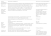
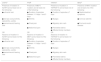
Table 1 shows the allergic and associated manifestations in different IEI.
Allergy manifestations and associated manifestations in different inborn errors of immunity.
| Allergy manifestations | Associated manifestations | Inborn errors of immunity(name/gene) |
|---|---|---|
| Elevated IgE and eosinophilia | Bacterial skin and pulmonary infections (with pneumatoceles);chronic mucocutaneous candidiasis;altered inflammatory response (cold abscesses);connective tissue abnormalities (hypermotility, scoliosis, retention of primary teeth, fractures, typical facies, aneurysms);non-flexural eczematous dermatitis; exanthema in the neonatal period. | STAT3 defect -Hyper-IgE S.ADa (Job S.)Defects in the STAT3 pathway - SNF341, IL6ST, IL6R |
| Atopy (atopic dermatitis, asthma)/food allergy/ anaphylaxis | Immune dysregulation; increased IgE and eosinophilia; viral skin infections; combined immunodeficiency (non-severe) | Actinopathies:Wiskott-Aldrich S. and defect in WIPb (thrombocytopenia with small platelets); defects in DOCK8, ARPC1B, CARMIL2Defects in CARD9, CARD11, CARD14, MALT1 |
| Increased IgE and eosinophilia; scoliosis;bacterial lung and skin infections;myoclonus and cognitive delay;lymphopenia. | PGM3 defect | |
| Immune dysregulation (enteropathy);bacterial / viral infections;short stature | STAT5b LFc | |
| Important eosinophilia; impaired growth. | JAK1 GFd | |
| Severe atopic dermatitis and/or erythroderma / ichthyosis | Eosinophilia | Omenn S. |
| Lymphoproliferation (hepatosplenomegaly, lymphadenopathy); severe combined immunodeficiency;barrier defects, with major ichthyosis; increased IgE and eosinophilia; bamboo hair; increased metabolic expenditure. | Comel-Netherton S. (SPINK5) | |
| Dermatitis with skin ulcers; increased IgE; bacterial skin and pulmonary infections; hepatosplenomegaly. | Prolidase deficiency | |
| Urticaria and anaphylaxis | Neonatal urticaria; very important eosinophilia; growth alteration. | STAT5b GFd |
| Early and severe hives; erythema and pruritus; anaphylaxis. | PLAID e (evaporative cooling; PLCG2 GF d)Familial vibratory urticaria (ADGRE2)Hereditary alpha-tryptasemia (TPSAB1) |
Source: Milner, 2020.9,10
Findings that raise suspicion of a primary allergic disorder in the absence of other associated manifestations, such as infections or autoimmunity, are as follows: very early onset; more severe and/or atypical manifestations and/or associated allergic manifestations; family history of autoimmunity and/or repeated infections, even in the absence of allergy; presence of characteristics such as overall developmental delay, cognitive impairment, joint hypermobility, scoliosis, and others.9,10
AutoimmunityApproximately a quarter of patients diagnosed with an IEI have one or more manifestations of autoimmunity or inflammation.5 In fact, manifestations of autoimmunity can appear in virtually all IEI; however, they are more common in patients with a diagnosis of common variable immunodeficiency and in patients with some cell immunity defect.5 Early and/or multiple manifestations of autoimmunity should raise the suspicion of an IEI as a underlying disease, especially if associated with infectious conditions.11,12
The autoimmune diseases most commonly associated with IEI are cytopenias: anemia, thrombocytopenia, neutropenia, and Evans syndrome (anemia and thrombocytopenia). The risk of autoimmune cytopenia is assumed to be approximately 120 times higher in individuals with an IEI than in the general population.5 Other manifestations of autoimmunity/inflammation that may be part of the clinical picture of IEI include the following: endocrinopathies, systemic lupus erythematosus, rheumatoid arthritis, juvenile idiopathic arthritis, vitiligo, bullous pemphigoid, atrophic gastritis, inflammatory bowel disease, and autoimmune enteropathy.13,14 Children diagnosed with an IEI are 80 times more likely to have inflammatory bowel disease and 40 times more likely to have arthritis than the general pediatric population.5
There are at least five clinical patterns of autoimmunity in IEI: “ALPS-like” (autoimmune lymphoproliferative syndrome)–autoimmune cytopenias and lymphoproliferation; “CVID-like” (common variable immunodeficiency)–hypogammaglobulinemia, infections, and hematological and solid organ autoimmunity; “IPEX-like” (enteropathy, autoimmune endocrinopathies, eczema, vasculitis); “IBD” (inflammatory bowel disease); and rheumatological diseases–Behçet, lupus, and monogenic juvenile idiopathic arthritis.2,15
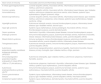
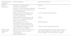
Table 2 describes the IEI that most often present with autoimmunity/inflammation and the associated clinical conditions.13
Inborn errors of immunity and most commonly related autoimmune and inflammatory manifestations.
| Inborn errors of immunity | Associated autoimmune/inflammatory diseases |
|---|---|
| X-linked agammaglobulinemia | Juvenile idiopathic arthritis, rheumatoid arthritis, inflammatory bowel disease, type I diabetes mellitus, autoimmune cytopenias |
| Common variable immunodeficiency | Juvenile idiopathic arthritis, rheumatoid arthritis, inflammatory bowel disease, type I diabetes mellitus, autoimmune cytopenias, pernicious anemia, celiac disease, systemic lupus erythematosus, Sjögren syndrome, vitiligo |
| Selective IgA deficiency | Juvenile idiopathic arthritis, rheumatoid arthritis, inflammatory bowel disease, type I diabetes mellitus, autoimmune cytopenias, celiac disease, systemic lupus erythematosus, Sjögren syndrome, vitiligo |
| HyperIgM syndrome | Autoimmune hemolytic anemia, immune thrombocytopenic purpura, inflammatory bowel disease, type I diabetes mellitus, discoid lupus |
| Hyper IgE syndrome | Systemic lupus erythematosus, bullous pemphigoid, rheumatoid arthritis, immune thrombocytopenic purpura, juvenile dermatomyositis |
| Omenn syndrome | Hashimoto’s thyroiditis, inflammatory bowel disease, immune thrombocytopenic purpura |
| DiGeorge syndrome | Immune thrombocytopenic purpura, autoimmune hemolytic anemia, Hashimoto’s thyroiditis, Graves’ disease, inflammatory bowel disease, uveitis, juvenile idiopathic arthritis |
| Wiskott-Aldrich syndrome | Immune neutropenia, immune thrombocytopenic purpura, autoimmune hemolytic anemia, rheumatoid arthritis, juvenile idiopathic arthritis, inflammatory bowel disease |
| Telangiectasia ataxia | Autoimmune hemolytic anemia, immune thrombocytopenic purpura, Hashimoto's thyroiditis, juvenile idiopathic arthritis |
| Autoimmune polyendocrinopathy candidiasis ectodermal dystrophy (APECED) | Autoimmune hypoparathyroidism, autoimmune hepatitis, Hashimoto's thyroiditis, Graves’ disease, primary cholangitis, type I diabetes mellitus, vitiligo, psoriasis |
| Immunodysregulation polyendocrinopathy enteropathy X-linked (IPEX) | Autoimmune enteropathy, type I diabetes mellitus, autoimmune cytopenias, Hashimoto’s thyroiditis, eczema |
| LRBA or CTLA4 deficiency | Autoimmune cytopenias, Hashimoto’s thyroiditis, inflammatory bowel disease, type I diabetes mellitus, systemic lupus erythematosus, juvenile idiopathic arthritis |
| Autoimmune lymphoproliferative syndrome (ALPS) | Immune thrombocytopenic purpura, autoimmune hemolytic anemia, systemic lupus erythematosus, rheumatoid arthritis, juvenile idiopathic arthritis |
| Chronic granulomatous disease | Systemic lupus erythematosus, inflammatory bowel disease, immune thrombocytopenic purpura, type I diabetes mellitus, juvenile idiopathic arthritis, rheumatoid arthritis |
| Complement system defects | Systemic lupus erythematosus, dermatomyositis, rheumatoid arthritis, juvenile idiopathic arthritis |
Adapted from Amaya-Uribe et al., 2019.13
Inflammatory conditions related to IEI present as hemophagocytic lymphohistiocytosis (HLH) or as autoinflammatory diseases.
HLH is a severe and often fatal hyperinflammatory syndrome associated with tissue infiltration (bone marrow, liver, ganglia, spleen, skin, and central nervous system) and high production of inflammatory cytokines by activated lymphocytes and macrophages.16,17 When related to autoinflammatory, autoimmune diseases, or malignancies, it is often called macrophage activation syndrome.18
Virtually any IEI in which immune dysregulation mechanisms are involved can evolve with an HLH, whether or not associated with viral infections, particularly Epstein-Barr virus (EBV). However, the defects classically related to this clinical condition are familial hemophagocytic lymphohistiocytosis (FHLH) syndromes with or without hypopigmentation.1,19 In both groups, there are cytotoxicity defects and the trigger for HLH may be a viral infection. Among the FHLH syndromes, defects in the PFR1 (perforin) and UNC13D genes are the most common.20 In hypopigmentation syndromes, Chédiak–Higashi syndrome, Griscelli syndrome type 2, and Hermansky–Pudlak syndrome types 2 and 10, there is partial oculocutaneous albinism, neurological disorders, neutropenia, and/or coagulation disorders.1
Cytotoxicity is normal or only partially affected in other IEI associated with HLH. IEI with EBV-related lymphoproliferation may also evolve with HLH: X-linked lymphoproliferative syndromes, ITK and CD27 deficiency, and XMEN syndrome (X-linked immunodeficiency with magnesium defect, Epstein-Barr virus infection, and neoplasia).21 Despite T-cell deficiency, combined (non-severe) immunodeficiencies can be complicated with HLH by macrophage activation.22 This mechanism is also responsible for HLH in autoinflammatory diseases, as in the NLRC4 defect, the monogenic autoinflammatory disorder characteristically associated with macrophage activation syndrome.17,18
Regarding IEI, the differential diagnosis with sepsis is crucial and, therefore, a high level of suspicion is necessary. What is identified is a condition similar to sepsis, but without the identification of an infectious agent and/or without improvement with antimicrobial use.21
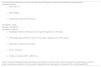
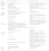
Table 3 describes the diagnostic criteria for HLH, based on the HLH-2004 protocol, updated in 2007.17,23 It is important to mention that ferritin is usually above 2000 μg/L and that values >10,000 μg/L have high diagnostic specificity in children.19,21 The only identified variable that is associated with higher mortality in HLH in patients with IEI is hypoalbuminemia. One study found that albumin values <3.07 g/dL were associated with a 5.8-fold increase in HLH mortality.22
Criteria for the diagnosis of hemophagocytic lymphohistiocytosis, primary or secondary.
| Five of the eight criteria below or clinical data with genetic diagnosis of disease associated with hemophagocytic lymphohistiocytosis |
|
|
|
| Hemoglobin <9 g/dL |
| Platelets <100,000/mL |
| Neutrophils <1000/mL |
|
|
|
|
|
| Other clinical and laboratory findings that may be associated: meningoencephalic signs and symptoms, adenomegaly, jaundice, edema, exanthema, alterations in liver enzymes, hypoproteinemia, hyponatremia, increased VLDL, and decreased HDL. |
VLDL, very low-density lipoprotein; HDL, high- density- lipoprotein.
The term autoinflammatory diseases (AID) was introduced in the late 1990s, to describe clinical conditions characterized by spontaneous inflammation in the absence of autoimmunity. Currently, it is known that AID constitute innate immunity regulation defects, in which there is hypersecretion of inflammatory cytokines. These diseases are characterized by a recurrent or persistent inflammatory process, in the absence of an infectious process and absence of autoantibodies or autoreactive T cells.24
In this text, we describe the monogenic AID, which appear in childhood as fever and clinical and laboratory signs of systemic inflammation, different skin rashes, and different patterns of sterile inflammation in other organs, which vary according to the specific autoinflammatory disease. The signs and symptoms are often recurrent and characteristic of each patient, most commonly involving skin and mucosal membranes, digestive tract, musculoskeletal system, and eyes.25
Considering Facing recurrent episodes of inflammatory manifestations, with or without fever, with laboratory evidence of altered inflammatory activity, it is important to attempt to identify any infectious agent or search for laboratory evidence of autoimmunity or malignant diseases. After ruling out these causes, the possibility of an AID must be considered, but the definitive diagnosis is only possible through genetic investigation. Aiming to better guide this investigation or to implement treatment in the absence of a genetic diagnosis, it is essential to characterize the clinical phenotype: age at symptom onset, ethnicity of ascendants, habitual triggers of episodes, duration and interval of acute episodes, and details on the signs and symptoms that characterize the condition.26,27
There have been many proposed classifications and they generally use clinical data or pathophysiological mechanisms. Table 4 shows a very useful classification, based on clinical data: fever patterns, type of skin lesion, and type of organ-specific inflammation.28
Autoinflammatory diseases classified according to fever pattern and/or type of skin lesion and associated inflammatory manifestations.
| Clinical manifestations | Autoinflammatory diseases |
|---|---|
| Group 1 | |
| Recurrent/episodic fever with or without skin rash | |
| Short-duration fever | Familial Mediterranean fever, Hyper IgD |
| Longer-lasting fever | TRAPS |
| Group 2 | |
| Neutrophilic urticaria | |
| Short-duration recurrent fever | FACS |
| Persistent inflammation with episodes of exacerbation | Muckle-Wells Syndrome, NOMID/CINCA |
| Group 3 | |
| Pustular exanthema and episodic fever | |
| Pyogenic disease with sterile osteomyelitis | DIRA, Majeed Syndrome |
| Pyogenic disease with sterile pyogenic arthritis | PAPA |
| Pustular disorder with a picture similar to Behçet | HA20 |
| Pustular disorder with psoriasis-like condition | DITRA, CAMPS, AMPS |
| Pustular disorder with inflammatory bowel disease | IL10, IL10R, NISBD1 |
| Pyogenic disease with varied mechanisms | PAAND, PFIT |
| Group 4 | |
| Vasculopathy and panniculitis/lipodystrophy | |
| Mediated by type I interferon | CANDLE, PRAAS |
| Partially dependent on TNF | ORAS |
| Group 5 | |
| Vasculopathy with or without vasculitis and livedo | |
| No significant demyelination and with interstitial pulmonary disease | SAVI |
| With central nervous system demyelinating disease | AGS. Pseudo TORCH |
| With spondyloenchondrodysplasia | SPENCD |
| With stroke | DADA2 |
| Group 6 | |
| Cutaneous granulomatosis | |
| Without immunodeficiency | Blau syndrome |
| With immunodeficiency | PLAID, APLAID, NDAS |
| Group 7 | |
| Macrophage activation syndrome | NLRC4, LACC1 |
| With NK and TCD8 defects and immunodeficiency | FLH, Chédiak-Higashi, Griscelli, Hermansky-Pudlak |
| Group 8 | |
| Others | Cherubism, SIFD, AISLE, NLRP12, TNFRSF11A, NAIAD |
TRAPS, TNF receptor-associated periodic syndrome; FACS, familial cold autoinflammatory syndrome; NOMID/CINCA, neonatal-onset multisystem inflammatory disease/ chronic infantile neurological cutaneous and articular syndrome; DIRA, deficiency of the interleukin-1 receptor antagonist; PAPA, pyogenic arthritis, pyoderma gangrenosum and acne syndrome; HA20,haploinsufficiency of A20; DITRA, deficiency of the IL-36 receptor antagonist; CAMPS, caspase activation and recruitment domains (CARD)14-mediated psoriasis; AMPS,AP1S3-mediated psoriasis; NISBD1,neonatal inflammatory skin and bowel disease 1; PAAND, pyrin-associated autoinflammation with neutrophilic dermatosis; PFIT, periodic fever, immunodeficiency, and thrombocytopenia; CANDLE, chronic atypical neutrophilic dermatoses with lipodystrophy and elevated temperature syndrome; PRAAS, proteasome-associated autoinflammatory syndromes; ORAS, otulin-related autoinflammatory syndrome; SAVI, stimulator of IFN genes (STING)-associated vasculopathy with onset in infancy; AGS, Aicardi-Goutières syndrome; SPENCD, spondyloenchondrodysplasia with immune dysregulation; DADA2,deficiency of adenosine deaminase 2; PLAID, cold-induced urticaria and or granulomatous rash; APLAID, PLCγ2 associated antibody deficiency and immune dysregulation; NDAS, nuclear factor (NF)-κB essential modulator (NEMO) deleted exon 5 autoinflammatory syndrome– X-linked; LACC1, LACC1-mediated monogenic Still disease; FLH, familial hemophagocytic lymphohistiocytosis; SIFD, congenital sideroblastic anemia, B-cell immunodeficiency, periodic fevers, and developmental delay; AISLE, autoinflammatory syndrome-associated with lymphedema; NAIAD, NLRP1-associated autoinflammation with arthritis and dyskeratosis.
Adapted from Goldbach-Mansky & de Jesus, 2019.28
Due to their frequency among the AID, Table 5 shows the new classification criteria proposed by the Eurofever project for hereditary recurrent fevers.29 The criteria proposed by Eurofever for periodic fever, aphthosis, pharyngitis, and adenitis (PFAPA)include at least seven among the following eight items: presence of pharyngotonsillitis; duration of episodes between three to six days; cervical adenitis; periodicity; with no diarrhea, chest pain, skin rash, or arthritis.29
Criteria for the classification of recurrent hereditary fevers.
| CAPSa | FFMb | TRAPSc | MKDd |
|---|---|---|---|
| Presence of mutation in NLRP3 and at least one of the following: | Presence of MEFV mutation and at least one of the following: | Presence of mutation in TNFRSF1A and at least one of the following: | Presence of MKV mutation and at least one of the following: |
| ● urticarial rash | ● Duration of episodes between 1–3 days 1 and 3 days | ● Duration of episodes ≥7 days | ● Gastrointestinal symptoms |
| ● red eye (conjunctivitis, episcleritis, uveitis) | ● Arthritis | ● Myalgia | ● Cervical adenitis |
| ● sensorineural hearing loss | ● Chest pain | ● Migratory skin rash | ● Foot-and-mouth disease |
| ● Abdominal pain | ● Periorbital edema | ||
| ● Affected family members | |||
| OR | OR | OR | |
| Absence of mutation in NLRP3 and at least 2 of the following: | Absence of mutation in MEFV and at least 2 of the following: | Absence of mutation in TNFRSF1A and at least 2 of the following: | |
| ● urticarial rash | ● Duration of episodes between 1–3 days 1 and 3 days | ● Duration of episodes ≥7 days | |
| ● red eye (conjunctivitis, episcleritis, uveitis) | ● Arthritis | ● Myalgia | |
| ● sensorineural hearing loss | ● Chest pain | ● Migratory skin rash | |
| ● Abdominal pain | ● Periorbital edema | ||
| ● Affected family members |
Adapted from Gattorno, 2019.29
Lymphoid proliferation with adenomegaly and/or splenomegaly occurs in many IEI and is closely related to immune dysregulation mechanisms with or without viral infections, particularly EBV. In addition there is the possibility that it is caused by bacteria or intracellular fungi infections, which will not be discussed here.
In association with immune dysregulation, due to apoptosis or cytotoxicity defects, lymphoproliferation is usually associated with autoimmune manifestations, mainly autoimmune cytopenias, endocrinopathies or enteropathy, or is associated with hyperinflammatory conditions, such as hemophagocytic lymphohistiocytosis.1
Despite the overlapping of mechanisms related to immune dysregulation with or without viral infections, one or another basic lymphoproliferation mechanism predominates in some IEI. Common variable immunodeficiency, the most common symptomatic IEI, corresponds to the defect most often related to lymphoproliferation caused by immune dysregulation, with others being autoimmune lymphoproliferative syndrome (ALPS) and activated p110δ syndrome (APDS). In the latter, persistent EBV and/or cytomegalovirus (CMV) viremia is common.1
Defects with EBV-related lymphoproliferation, usually without autoimmunity manifestations, are the X-linked lymphoproliferative syndromes (caused by mutations in SAP or XIAP), and CD27, CD70, CTPS1, CD137, RASGRP1, RLTPR, PRKCD, and XMEN deficiencies.1
When treating a patient diagnosed with an IEI with lymphoproliferation, considering the possibility of infections and the associated malignancy potential, surveillance through ganglion biopsies, ideally by excision, with phenotyping and clonality studies, in addition to investigating infectious agents, are essential.30
The IEI that are most commonly related to lymphoproliferation are shown in Table 6.1
Inborn errors of immunity according to the lymphoproliferation mechanism and associated clinical and laboratory manifestations.
| Lymphoproliferation mechanism | Other manifestations | Inborn error of immunity |
|---|---|---|
| Immune dysregulation | Combined T and B cells defect, eosinophilia, erythroderma | Omenn S. |
| Combined T and B cells defect, progressive CD4 lymphopenia, normal B cells, normal to low immunoglobulins | ITK deficiencya | |
| Antibody defect, decreased IgG and IgA and/or IgM, sinopulmonary infections, autoimmune cytopenias | Common variable immunodeficiency | |
| Antibody defect, decreased IgG and IgA with normal or increased IgM, bacterial infections, autoimmunity | APDSa,b | |
| Antibody defect, low or normal immunoglobulins and B lymphocytes, hematological and thyroid autoimmunity | NFKB1 deficiencya | |
| Treg cell defects, hematological or solid organ autoimmunity, enteropathy | CD25 and CD122a deficiencySTAT3GFc | |
| Related to EBV (Epstein-Barr virus) | Normal or high T cells, normal or reduced memory B cells, normal or reduced immunoglobulins, dysgammaglobulinemiad | X-linked lymphoproliferative syndromes (by mutations in SAP or XIAP), deficiencies in CD27, CD70e, CTPS1, CD137, RASGRP1, RLTPR, PRKCDe and XMEN |
Source: Tangye, 2020.1
Malignancies occur more frequently and earlier in life in patients with IEI and are the second cause of death in children and adults with this diagnosis, after infectious conditions.31,32 The increased risk of cancer in patients with IEI identified by the USIDNET study6 refers mainly to lymphomas, leukemias, and stomach cancer.
One of the immune system’s functions is to identify and eradicate tumor cells, known as immune surveillance. Many immune system pathways that protect against infections also act in this surveillance.33 However, the risk of the most common malignancies in the general population of men and women (colon, breast, lung, and prostate) is similar in individuals with IEI.6 This means that the immune surveillance system probably plays a limited role in the control of these tumors.33
Considering the limited spectrum of malignancies associated with IEI, the small proportion of patients with IEI among patients with malignant diseases, and the relatively small number of IEI related to cancer, other mechanisms besides immunological surveillance have been implicated.31 Other mechanisms proposed for the association between IEI and malignancies include the following: chronic tissue inflammation, DNA repair defects, telomere maintenance defects, myeloid cell development defects, infections, as well as apoptosis, cytotoxicity or metabolic defects.31 Many malignancies in patients with IEI are related to viral diseases, most commonly EBV, which appears to be related to the development of 30% to 60% of lymphoma cases.34 However, other viruses may be involved: the human herpes virus (HHV 6 and 8), the human papillomavirus (HPV), the human T-cell lymphotropic virus (HTLV), and CMV.35,36
The most common malignancies identified in patients with IEI are non-Hodgkin's lymphoma, mainly the B-cell type, leukemias, and gastric cancer. They affect patients with antibody defects, especially with common variable immunodeficiency, due to their high frequency among the IEI.33,36 However, the IEI with the highest risk of evolution to a malignant disease is ataxia-telangiectasia, in which there is a defect in DNA repair.34Regarding solid tumors, the one most commonly described in patients with IEI is the gastric carcinoma, particularly in common variable immunodeficiency .34
IEI patients tend to have malignancies earlier in life than the general population. One study shows that lymphomas appear between 7 months and 76 years, with a mean age of 12 years.36 The chance of a child with an IEI to develop cancer varies between 5% and 25%, and the type of cancer depends on the type of immune defect.37
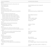
Table 7 shows the IEIs most commonly related to the development of cancer and the malignancies most often associated with them.33,38
Inborn errors of immunity most often related to the development of cancer, type of defect involved, and associated malignancies.
| Types of defects | Inborn errors of immunity | Associated malignancies |
|---|---|---|
| Disorders with DNA break | Ataxia telangiectasia | Lymphomas and leukemias, breast cancer |
| Bloom syndrome | Lymphomas, acute leukemias and carcinomas | |
| Nijmegen syndrome | Lymphomas, leukemias, CNS tumor | |
| DNA ligase IV mutations | Lymphomas, leukemias | |
| Antibody defects | Common variable immunodeficiency | Non-Hodgkin's lymphoma, gastric, thyroid or skin cancer. |
| Activated p110δ syndrome(APDS) | Lymphomas | |
| Selective IgA deficiency | Gastrointestinal tract tumor | |
| Non-severe combined defects(T and B) | Artemis syndrome, ADA defects, ZAP 70, RAG1 and Coronin 1A (non-severe phenotype) | Lymphomas and carcinomas |
| Wiskott-Aldrich syndrome | Lymphomas, acute lymphocytic leukemia, Kaposi's sarcoma, myelodysplastic syndrome | |
| Hair cartilage hypoplasia | Lymphomas | |
| DOCK8 mutation | Lymphomas, leukemias, epithelial tumors, and others | |
| DiGeorge syndrome | Lymphomas and leukemias, thyroid carcinoma, neuroblastoma, hepatoblastoma, Wilms' tumor. | |
| Hyper IgM syndrome | Liver, pancreas, and biliary pathway carcinomas | |
| Immune dysregulation | Autoimmune lymphoproliferative syndrome (ALPS) | Hodgkin's and non-Hodgkin's lymphomas |
| Susceptibility to EBV:X-linked lymphoproliferative syndrome, deficiencies of ITK,CD70, CD27, RASGRP1, CTPS1, CD137, MAGT1, CARMIL2, PRKCD | EBVa-associated lymphomasa | |
| Familial hemophagocytic lymphohistiocytosis associated with hypopigmentation | Lymphomas | |
| IL10R deficiency | Lymphomas | |
| Chronic mucocutaneous candidiasis | Squamous cell tumors of the oral cavity and esophagus | |
| Bone marrow defects | GATA2 deficiency | Myelodysplastic syndrome, acute myeloid leukemia, chronic myelomonocytic leukemia, EBVa and HPVb tumors |
| Fanconi anemia | Myelodysplastic syndrome, acute myeloid leukemia, squamous cell carcinomas of the head and neck, breast cancer | |
| Congenital dyskeratosis | Leukemias, myelodysplastic syndrome, squamous cell carcinomas of the head and neck, lung tumors, gastrointestinal tract, and liver | |
| Mirage syndrome and ataxia-pancytopenia syndrome | Myelodysplastic syndrome and acute myeloid leukemia | |
| Congenital neutropenia | Myelodysplastic syndrome and acute myeloid and acute lymphocytic leukemia | |
| Shwachman-Diamond syndrome | Acute or chronic myeloid leukemias | |
| Defects in innate immunity | Verruciform epidermodysplasia | Skin cancer (HPV)b |
| WHIMc | Skin cancer |
Source: Mortaz, 2016; Haas, 2019; Kebudi, 2019; Riaz, 2019; Renzi, 2020; Rezaei, 2020; Khalil, 2020.33-38
The treatment of non-infectious manifestations of IEI is, at least initially, similar to that performed for these same manifestations when they occur outside the context of IEI. The use of immunosuppressive medications requires extra care, as there is a risk of increasing the chance of infections.39
However, in more severe cases and/or poorly responsive to these therapeutic resources, the diagnosis of a monogenic disease implies the indication of drugs directed to the immune system pathways involved in the specific defect and/or hematopoietic stem-cell transplantation (HSCT).2,39 One example is the very early-onset inflammatory bowel disease related to a monogenic defect: when severe and/or unresponsive to immunosuppressants or immunobiological agents, HSCT is indicated and is curative. Gene therapy is a prospect for the perhaps not-too-distant future.39
ConclusionsNot only should repeated, severe, difficult-to-treat, and/or opportunistic microorganism infections raise suspicion of IEI, but also manifestations of allergy, inflammation, autoimmunity, lymphoproliferation, or cancer, particularly if they are recurrent, associated with each other, affect young patients, present as severe conditions, and/or are difficult to treat.
The initial treatment of non-infectious manifestations associated with IEI differs little from their treatment when they are not related to IEI. However, when an IEI is diagnosed, specific medications may be indicated and, in severe cases, hematopoietic stem cell transplantation appears as a therapeutic option.
Conflicts of interestThe authors declare no conflicts of interest.


