

Evaluate autonomic function and low-grade inflammation and characterize the correlation between these variables in schoolchildren with obesity living in the Brazilian northeast region.
Methods84 children with obesity and 41 with normal weight were included in this cross-sectional study. Anthropometry, body composition, blood pressure (BP), inflammatory biomarkers, and heart rate variability (HRV) indexes were analyzed in children aged 7 to 11 years.
Resultschildren with obesity had increased systolic (p = 0.0017) and diastolic (p = 0.0131) BP and heart rate (p = 0.0022). The children with obesity displayed significantly lower SDNN, RMSSD, NN50, HF (ms), HF (nu), SD1, SD2, and higher LF (ms), LF (nu), LF/HF, SD1/SD2, DFA-α1, and DFA-α2, compared to normal weight. A lower and higher capacity for producing IL-10 (p = 0.039) and IL-2 (p = 0.009), respectively, were found in children with obesity compared to children with normal weight. Although IL-2, IL-4 and IL17A did not correlate with HRV parameters, IL-6 was positively correlated with SDNN, LF (ms) and SD2, TNF-α was positively correlated with LF/HF and SD1/SD2 ratio, and IFN-γ was positively correlated with SDNN, RMMSSD, NN50, LF (ms), HF (ms), SD1, and SD2.
ConclusionsThe findings suggest that children with obesity have impaired autonomic function and systemic low-grade inflammation compared to children within the normal weight range, the inflammatory biomarkers were correlated with HRV parameters in schoolchildren living in the northeastern region of Brazil.
Childhood obesity is a global health issue associated with infancy and lifelong adverse health consequences, including an increased risk of suffering or developing cardio-metabolic diseases.1 In Brazil, the prevalence of childhood obesity in the last three decades was 8.2%, increasing with age, decade, and in more developed regions.2
Traditional factors, such as insulin resistance, hypertension, and reduced high‐density lipoprotein cholesterol (HDL‐C) are linked to cardiovascular disease. However, the detection of the early stages of cardiac autonomic dysfunction and related causes could be a useful strategy to identify the presence of cardiovascular risk in childhood obesity, and heart rate variability (HRV) could be used as a warning sign of cardiac diseases.3
Autonomic nervous system (ANS) control of the heart is a dynamic process modulated by parasympathetic and sympathetic innervation.4 RR-variation or HRV obtained from an electrocardiogram, Holter monitor or chest strap has been a commonly used and validated test to assess cardiac autonomic function through time, frequency, and non-linear domains indices that quantify parasympathetic (RMSSD, NN50, HF, and SD1), and sympathetic (LF, LF/HF and SD2) modulation.5
Although an early study carried out with German children has not found significant differences in HRV parameters between children with normal weight and obesity,6 cardiac autonomic dysfunction and reduced HRV has been reported in children with obesity.7,8 Concomitantly, chronic low-grade inflammation and obesity have been reported in children.9 In this condition, excess adipose tissue dysregulates the immune system, affecting the balance and levels of cytokines and increasing the levels of pro-inflammatory interleukin-6 (IL-6), tumor necrosis factor-alpha (TNFα), and C-reactive protein (CRP).10,11
Regarding the association of autonomic impairment with chronic low-grade inflammation in children with obesity, The authors found only one cross-sectional study with a reduced sample size demonstrating that impaired HRV and low-grade chronic inflammation coexist in childhood obesity in the United States of America, but no significant relationships were found between any of the measured HRV variables and cytokines levels.9 Considering that inflammation and autonomic function may be modulated by the environment and lifestyle and may have ethnic-specific implications, and the scarcity of studies linking inflammation and autonomic function in children, this study aimed to evaluate autonomic function and low-grade inflammation and to characterize the correlation between these variables in schoolchildren with obesity living in the Northeastern region of Brazil. The hypotheses tested are that children with obesity have lower HRV and higher low-grade inflammation compared to normal-weight children and that there is a positive association between autonomic dysfunction and chronic low-grade inflammation in children living in the Northeast region of Brazil.
MethodsEthical aspectsThis cross-sectional study was conducted in agreement with the declaration of Helsinki. The study was approved by a Human Research Ethics Committee (Health Sciences Center, Federal University of Paraiba – João Pessoa – PB – Brazil; Reference number protocol nº 4.676.103) and all procedures were conducted in agreement with Resolution 466/2012 of the National Health Council. All parents received information about the study and gave written informed consent before data collection.
Participants and study designAccording to data from the Department of Education and Culture of the Municipality of João Pessoa (SEDEC), in 2020 there were 23.861 students regularly enrolled in 97 primary public schools in the municipality of João Pessoa (PB, Brazil), distributed in the four geographic regions of the municipality (north, south, east and west). To calculate the sample size, the following parameters were considered: the size of the reference population equal to 23.861 children from the 1st to the 5th year of primary public schools, the prevalence of obesity outcome equal to 13.3%,12 95% confidence interval, test power of 80%, and design effect (deff) equal to 1. Based on these parameters, the minimum size of the original sample was established at 76 children, a number that was plus 20% to compensate for losses and refusals, resulting in a final sample of 125 children.
Children between 7 and 11 years of age, of both sexes, enrolled in public primary schools were included in the study, and recruitment and data collection were conducted in schools. Children were excluded if they presented any of the following: physical inability to perform the anthropometric measures, psychological or behavioral disturbances, use of any medication, or having a disease compromising any analysis.
In the study, 125 children were allocated to two groups: 84 children in the obesity (OB) group and 41 in the normal weight (NW) group. Children were evaluated for anthropometry, body composition, and cardiovascular parameters, evaluating blood pressure, HRV, and cytokines levels. All collections were performed at the school.
Anthropometry and body compositionThe body weight was measured with an electronic scale (Omron®, HBF-514C) and height was assessed using a stadiometer (alturaexata®). Nutritional status, using body mass index for age (BMI/A) and sex according to WHO standards was determined using Anthor plus (version 1.0.4; WHO) Children were classified in normal weight: ≥ −2 z-score ≤ +1, overweight: > +1 z-score ≤ +2, and obesity z-score > +2.
To measure the waist circumference, a flexible steel measuring tape with a scale of 0–200 cm and precision of 0.1 mm (Sanny®) was used. Skinfolds were measured three times always on the right side of the body, using a scientific adipometer with a precision of 0.1 mm (Sanny®). The average values were calculated and subsequently used to estimate the body fat percentage.
Blood pressure measurementBlood pressure was measured with a digital sphygmomanometer (Omron healthcare® HBP-1100) validated for use in children by the Brazilian Society of Cardiology. After an initial stabilization period (5 min), three consecutive measurements were taken for each child at 1 min intervals, and the average values were used in the analysis. The measurement was performed with the child in a seated position and with an adequate cuff. In addition, the arm was free and positioned at heart level, resting on a surface and with the palm facing up.
Heart rate variability analysisIt was recommended that children do not engage in vigorous physical activity or drink caffeinated beverages for at least 24 h prior to HRV analysis. Recordings were made in the morning in a quiet room. A short-term recording was performed with the children at rest, in the supine position, and breathing normally at tidal volume. HRV analysis was performed in 10 min record, according to previous studies.13-15 A smartphone (Iphone12) was used to record the RR intervals on an APP (Elite HRV LLC, Asheville, NC, USA, and Release 4.0.2, 2018). An elastic belt of adjustable size was positioned comfortably around the participant's chest and a transmission device (Polar model H10, Polar Electro, Finland; 5 kHz transmission system coded with Owncodes, Polar Electro) was attached on the front at the level of the xiphoid process. The raw data recorded was exported as a text file and analyzed by Kubios HRV Standard software (version 3.2.0; Biosignal Analysis and Medical Image Group, Department of Physics, University of Kuopio, Kuopio, Finland). The interpolation of the series was performed by cubic spline, and frequency values of 4 Hz were set. The R-wave time instants were automatically detected by applying a built-in QRS detection algorithm of Kubios software and when ectopic beats exceeded 5% of the total analyzed, the measurement was repeated. Early studies have demonstrated that smartphone apps and chest strap (Polar) provide excellent electrocardiogram compliance for all variables in the time domain, frequency domain, and nonlinear indexes.16,17 The following measures of HRV analysis were determined; 1) time-domain parameters: the standard deviation between the duration of NN intervals (SDNN), the square root of the mean of the sum of the squares of the successive differences between adjacent normal-to-normal beats (RMSSD), adjacent NN with differences in duration greater than 50 (NN50) low-frequency band (LF), high-frequency band (HF), standard deviation of the instantaneous variability of continuous NN intervals in the Poincaré graph (SD1), standard deviation of long-term continuous NN intervals (SD2) and detrended fluctuation analysis (DFA).
Blood samples and cytokines measurementsBlood samples were collected by a qualified nurse after 12 h fasting and without strenuous exercise in the last 24 h. Serum samples were used for the measurement of cytokines through the Cytometric Bead Array (CBA) technique. Th1/Th2/Th17 CBA kits from Becton Dickinson Biosciences were used to quantify the cytokines IL-2, IL-4, IL-6, IL-10, IL-17a, IFN- γ, and TNF- α. The Becton Dickinson (BD)™ CBA uses a series of particles (microspheres or beads) with different fluorescence intensities to simultaneously detect soluble cytokines through a capture surface. Each bead is conjugated to a specific antibody and cytokines are detected by the fluorochrome phycoerythrin. The fluorescence intensity of each complex reveals the cytokine concentration (pg/mL). An Accuri C6 BD® flow cytometer was used, and analysis of CBA data was performed using FCAP 1.0.1 software.
Statistical analysisThe data normality was assessed by the Kolmogorov-Smirnov test. Descriptive statistics are presented as mean (standard deviation) or median (min-max). Comparison intergroup (NW and OB) was performed using Student's t-test in the case of the parametric data or the Mann-Whitney test for non-parametric data. The relationships between variables were analyzed using Pearson or Spearman correlation test according to data distribution. The statistical analysis was performed using GraphPad Prism® (version 6.01) and the significance was maintained at p < 0.05. An a posteriori power analysis was performed to assess differences between children with obesity and normal weight. Power was calculated using a two-sided 95% confidence interval, a prevalence of 63.51% of children with obesity having a heart rate above the mean, a prevalence of 35.48% of children with normal weight having a heart rate above the mean, a prevalence ratio of 1.8; and a prevalence difference of 28.03. The statistical power was performed using OpenEpi software (version 3.01) and the value obtained was 75.75%. Lastly, the effect size between‐groups differences were calculated using Cohen's d test and classified as small (d ≥ 0.20 and < 0.50), medium (d ≥ 0.50 and < 0.80), and large (d ≥ 0.80).
ResultsA total of 1.957 children were evaluated and classified according to BMI, with 1.009 males and 948 females. Of the total number of children assessed, 79 (4.04%) were classified as underweight, 1.249 (63.82%) as normal weight, 328 (16.76%) as overweight, and 301 (15.38%) as obese. One hundred and twenty-five of these children were included in this study, 41 with NW and 81 with obesity.
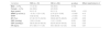
The anthropometric and cardiovascular characteristics of children included in the study are presented in Table 1. The groups were similar in terms of sex (p = 0.702) and age (p = 0.205). The obesity group had higher BMI/A (p < 0.0001), waist-hip ratio (WHR) (p < < 0.0001), body fat (BF) (p < 0.0001), SBP (p = 0.0017), DBP (p = 0.0131), and heart rate (HR) (p = 0.0022) when compared to the NW group, with the medium effect size for SBP, DBP and HR.
Anthropometric and cardiovascular characteristics of children with normal weight (NW) and obesity (OB).
Values are expressed as n (%), mean (SD) or median (min-max), and applied chi-square, T test or † Mann–Whitney, respectively.
BMIA, Body mass index for age; WHR, waist hip ratio; BF, body fat; SBP, Systolic blood pressure; DPB, Diastolic blood pressure; HR, heart rate.
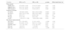
As reported in Table 2, children with obesity had increased IL-2 levels (p = 0.009) and decreased IL-10 levels (p = 0.039) when compared to the NW group (Table 2), with a medium effect size for the IL-2 and small effect size for the IL-10. Serum levels of IL-4, IL-6, IL-17A, TNFα, and IFN-γ were similar between groups (Table 2, p > 0.05).
Comparison of the values of cytokines between children with normal weight (NW) and obesity (OB).
Values are expressed as median (min-max). Mann-Whitney test.
IL-2, interleukin-2; IL-4, interleukin-4; IL-6, interleukin-6; IL-10, interleukin-10; IL-17A, interleukin-17A; TNF-α, tumor necrosis factor-α; IFN-γ, interferon-γ.
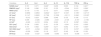
Regarding the HRV analysis, children with obesity had a predominance of sympathetic ANS in the detriment of parasympathetic. In the time domain, children with obesity had decreased SDNN (p = 0.002), RMSSD (p = 0.001), and NN50 (p = 0.008) compared to the NW children, with a larger effect size for SDNN and RMSSD (Table 3). In time-frequency, children with obesity had increased LFnu band (p = 0.001) and LF/HF ratio (p = 0.003) and decreased HFnu band (p = 0.004) compared to NW children, with the medium effect size for the three variables (Table 3). In non-linear indices, children with obesity had decreased SD1 (p < 0.000) and SD2 (p = 0.004), and increased SD2/SD1 ratio (p = 0.001), DFA-α1 (p = 0.002), and DFA-α2 when compared to NW children, with medium effect size for SD2/SD1 and DFA-α1 and larger effect size for DFA-α2 (Table 3).
Assessment of heart rate variability in children with normal weight (NW) and obesity (OB).
Values are expressed as mean (SD) or median (min-max); T test or † Mann–Whitney, respectively.
SDNN, standard deviation between NN intervals; RMSSD, square root of the mean of the square of the differences between consecutive NN intervals; NN50, adjacent NN with differences in duration greater than 50; LF, low frequency; HF, high frequency; SD1, standard deviation of the instantaneous variability of continuous NN intervals in the Poincaré graph; SD2, standard deviation of long-term continuous NN intervals; DFA, detrended fluctuation analysis; ms: milliseconds; nu: normalized units.
Correlations between HRV and inflammatory variables are shown in Table 4. Although IL-2, IL-4, and IL17A did not correlate with HRV parameters, IL-6 showed a positive correlation with SDNN, LF, and SD2, and IL-10 with LF (ms). TNF-α was positively associated with LF/HF ratio and SD1/SD2 ratio. IFN-γ showed a positive correlation with SDNN, RMSSD, NN50, LF, HF, SD1, and SD2.
Linear correlation coefficients between heart rate variability measures and inflammatory variables.
| Variables | IL-2 | IL-4 | IL-6 | IL-10 | IL-17A | TNF-α | IFN-γ |
|---|---|---|---|---|---|---|---|
| SDNN (ms)† | 0.161 | 0.071 | 0.246* | 0.206 | 0.033 | 0.134 | 0.401* |
| RMSSD (ms)† | 0.148 | 0.086 | 0.161 | 0.125 | −0.044 | 0.015 | 0.299* |
| NN50 (ms)† | 0.116 | 0.118 | 0.115 | 0.071 | −0.043 | 0.010 | 0.229* |
| LF (ms)† | 0.0856 | 0.0853 | 0.2924* | 0.2317* | 0.1037 | 0.1957 | 0.4713* |
| HF (ms)† | 0.1147 | 0.0600 | 0.1798 | 0.1528 | −0.0111 | 0.0061 | 0.3163* |
| LF (nu) | −0.012 | 0.024 | 0.088 | 0.048 | 0.155 | 0.166 | 0.113 |
| HF (nu) | 0.006 | −0.026 | −0.089 | −0.041 | −0.149 | −0.167 | −0.106 |
| LF/HF† | −0.023 | −0.010 | 0.034 | 0.023 | 0.115 | 0.239* | 0.024 |
| SD1 (ms)† | 0.134 | 0.083 | 0.161 | 0.136 | −0.041 | 0.010 | 0.308* |
| SD2 (ms)† | 0.151 | 0.062 | 0.256* | 0.217 | 0.052 | 0.167 | 0.425* |
| SD1/SD2 (ms)† | −0.071 | −0.034 | 0.029 | 0.019 | 0.173 | 0.219* | −0.024 |
| DFA-α1 | −0.030 | 0.036 | 0.118 | 0.077 | 0.162 | 0.196 | 0.139 |
| DFA-α2 | 0.033 | −0.037 | −0.068 | −0.093 | 0.010 | −0.006 | −0.177 |
SDNN, standard deviation between NN intervals; RMSSD, square root of the mean of the square of the differences between consecutive NN intervals; NN50, adjacent NN with differences in duration greater than 50; LF, low frequency; HF, high frequency; SD1, standard deviation of the instantaneous variability of continuous NN intervals in the Poincaré graph; SD2, standard deviation of long-term continuous NN intervals; DFA, detrended fluctuation analysis; ms: milliseconds; nu: normalized units.
This study identified that children with obesity had higher levels of SBP, DBP, HR, and higher serum levels of IL-2 and a lower serum level of IL-10 than NW children. The children with obesity also had autonomic dysfunction, as observed in linear and non-linear indices. The authors also found a positive correlation between HRV indices and inflammatory biomarkers, suggesting that autonomic and inflammatory dysfunction may occur simultaneously in children.
Some important variables related to obesity and cardiovascular disease were also different in these children, such as the increase in SBP, DBP, and HR. There is strong evidence of greater sympathetic activation in obesity, including changes in resting BP and HR, which is consistent with the results found in this study in children with obesity. It is important to highlight that, despite the existing differences according to nutritional status, the values of BP and HR were within the normal limits proposed by the Brazilian Guidelines of Hypertension and III Guidelines of the Brazilian Society of Cardiology on Analysis and Issue of Electrocardiographic Reports, respectively.18-20
Inflammation in obesity occurs due to functional changes in adipose tissue, resulting in the secretion of pro-inflammatory cytokines produced by activated macrophages residing in adipose tissue, and decreased secretion of protective adipokines, such as adiponectin.21 The mechanisms underlying this dysfunction occur in response to the structural changes in adipose tissue and may involve endoplasmic reticulum stress, hypoxia, and cellular senescence.21 Chronic low-grade inflammation results from a persistent failure to organize the inflammatory response. The interaction of macrophages and monocytes and other inflammatory mediators has been studied in the pathogenesis of obesity.22
This study was the first to evaluate a broad cytokine profile in children and showed that although IL-4, IL-6, IL17A, TNF-α and IFN-γ were similar between groups, children with obesity had increased serum levels of IL-2 and decreased serum levels of IL-10 compared to NW children, suggesting that children with obesity may have a loss of ability to regulate the monocyte-mediated immune response to produce cytokines.23 Physical inactivity can, among other things, lead to obesity, impair immune system function, and increase the risk of low-grade chronic inflammation. A higher concentration of IL-2-producing cells and a lower capacity to produce IL-10 have previously been reported in sedentary children compared to active children.24 It has been demonstrated that obesity alters immune function and that the imbalance between pro- and anti-inflammatory mediators in obesity may induce the establishment or progression of cardiovascular complications and dysautonomia.10,25
HRV is a measure of the natural changes occurring between beats in a row in heart rate, which represents the number of heart beats per minute.26 The HRV measurement must be carried out on normal cardiac cycles (N), without artifacts and ectopic beats. For this reason, The authors used the denomination N-N interval and not the R-R interval in this study.5 An optimal HRV level reflects adequate functioning, characterized by an inherent capacity for self-regulation, adaptability, or resilience. In contrast, a very small variation indicates age-related system depletion, chronic stress, pathology, or improper functioning at various levels of self-regulatory control systems.26
HRV analysis using linear and non-linear methods has been used to better understand complex and dynamic heart rhythms. In 2010, cross-sectional studies conducted in Brazilian children with obesity aged between eight and 12 years showed a reduction in non-linear HRV measures and decreased LF and HF indices, suggesting a reduction in both sympathetic and parasympathetic activities.27,28 Another study conducted in Brazilian children showed increased LFnu band and LF/HF ratio in children with obesity when compared to children within the normal weight range but without changes in non-linear HRV measures.8
The present study showed that children with obesity had decreased HRV measures in the time domain, increased LFnu and LF/HF ratio in the frequency domain, and increased SD2/SD1 and DFA-α1 and DFA-α2 in non-linear measures. The results may indicate unresolved cardiac stress characterized by parasympathetic withdrawal and sympathetic hyperactivation in obese children.29 Although it is not possible to determine the underlying causes involved in the fluctuations in HRV variables in children with obesity, it is reasonable to suggest that lifestyle changes over the last decade, particularly evidenced by the increase in consumption of ultra-processed and hypercaloric foods, associated with reduced physical activity, may have played a key role in altering HRV indices. This reaffirms the importance of the present study, which is essential to understand and identify the potential risk of autonomic dysfunction in children, as well as to propose effective intervention strategies to prevent cardiovascular disease later in life.
Regarding the contribution of hormonal, reflex, metabolic, endothelial, and inflammatory mechanisms in cardiovascular homeostasis mediated by the autonomic nervous system,18 the main mechanisms to explain alterations of the ANS in obesity are sympathetic overactivity, dysfunction of arterial baroreceptor properties, a key mechanism involved in the control of sympathetic and vagal tonus,18 and impairment of the respiratory-cardiovascular coupling, which may result in sympathetic activity and autonomic dysfunction.18
The positive correlations found between HVR indices in the time, frequency, and non-linear domains with biomarkers of inflammation are consistent with available evidence regarding the association between inflammation and ANS activity.18,30 An early study found a positive association between HRV and pro-inflammatory markers, although a meta-analysis had shown that a negative correlation between HRV and pro-inflammatory markers is more common.30 In this sense, an important point to highlight is the fact that vagal hyperactivity is positively correlated with first-response inflammatory mediators, such as TNF-α and IFN-γ, through the elimination of the parasympathetic nervous system via acetylcholine, which may partially explain the positive correlation of RMSSD, NN50, HF and SD1 with IFN-γ. On the other hand, the positive correlation of LF and SD2 with IL-6 indicates a slower action, characteristic of the sympathetic nervous system via epinephrine and norepinephrine. Furthermore, parameters that reflect total HRV (e.g., SDNN) are influenced by both mechanisms, which explains the positive correlation with IL-6 and IFN-γ.30
This study contributes to a better understanding of dysautonomia and low-grade chronic inflammation in children, demonstrating the coexistence of these conditions in children with obesity from the Northeast region of Brazil. The positive correlation between HRV indices and inflammation provides valuable information and encourages mechanistic studies to better elucidate this issue. In addition, this study may help to establish reference values for HRV indices in children, which could be an essential screening tool for cardiovascular risk in health outpatient clinics.
This study has some limitations. First, the cross-sectional design allows only the study of associations between variables, not causal relationships. Second, HRV and inflammatory biomarkers may differ by sex, and stratification by sex could be examined in further studies; and third, a broad assessment of metabolic variables panel could help to better understand the cardiometabolic risk in children with obesity in a post-pandemic scenario. These limitations will be addressed in future studies.
ConclusionThe results of this study show several cardiovascular risk factors in obese children compared to children in the normal weight range. At such a young age, obesity is already associated with lower HRV and low-grade chronic inflammation, which reflects and reinforces the need to develop effective strategies to prevent and combat childhood obesity. Furthermore, the correlations between inflammatory markers and HRV indices underscore the importance of elucidating the mechanisms by which such interactions may occur.
This study was funded by Paraiba State Research Foundation (FAPESQ, grant# 41903.612.28794.22092020, Edital nº005/2020) and CAPES (Finance code 001). de Brito Alves, JL thanks CNPq for their research productivity fellowship.


