
To analyze bone mineral content (BMC) and area bone mineral density (aBMD) accrual in adolescent male footballers who started their first football season.
Methods17 athletes (14.8 ± 0.4 years) were monitored across 15 weeks of football training. Participants were evaluated for somatic maturation (HPHV), BMC, and aBMD at three time points: before (M1) and after (M2) a preparatory phase, and at the end of the competitive phase (M3). BMC and aBMD were measured using DXA scans. Participants were divided into groups according to maturation status (circa-PHV and post-PHV), and the amount of accumulated training load (median split).
ResultsA significant effect (12.1 g/week, standard error (SE) = 2.6 g/week) was observed for lower limbs BMC across the three time points. There were no significant effects of time for upper limbs BMC. There was a significant effect of time for total body aBMD (0.007, SE = 0.003 g/cm2/week) across the three time points. Adolescents at post-PHV had a significant 245.6 g (SE = 56.1 g) higher BMC compared to adolescents at circa-PHV. No significant effects were observed for the accumulated training load.
ConclusionSystematic football training, even during the growth spurt, has a positive impact on adolescent bone markers despite the accumulated training load and maturation.
During human growth, skeletal development unfolds through the coordinated process of bone deposition and remodeling, leading to the adult skeleton as a result of the periosteal apposition of cortical bone and endochondral ossification.1 It is well known that adolescence is the most important period for bone mineral content accumulation, with bone mineralization increasing by ∼40%.2 This bone mass accrual during adolescence is attributed to the peak of height velocity and the peak of bone mass accrual, indicators of maximum growth in stature and greatest bone mass accrual, respectively.3,4 Baxter‐Jones, et al.5 demonstrated that ∼39% of total young-adult bone mass is acquired during the five-year period around peak-height velocity (PHV), and 95% of total bone mineral content is achieved up to 4 years after PHV.5 These changes in bone structure and composition occur at puberty and contribute to bone strength, resulting in a decreased risk of osteoporosis later in life.6
Among the main determinants of bone mass gains during adolescence are the modifiable factors such as nutritional intake and the mechanical load on the skeleton,7,8 which can explain 20–40% of the bone mass observed in adulthood. Importantly, a study using computer simulations indicated that increasing bone mineral by >10% of the mean observed in the first decades of life delays the onset of osteoporosis by 13 years. Being physical exercise and activity are the main causes of mechanical load in the skeleton,9 efforts are in place to increase the current understanding of the impact of different physical exercises on the bone health of adolescents, including participation in different sports.
Sports participation has been shown to be the most frequent manifestation of exercise in the pediatric population.10 However, considering the specific patterns of mechanical stimulus on the skeleton, each sport can differently affect the bone tissue at each body site.11 For example, basketball and gymnastics are defined as high-impact modalities because of the number of jumps during the practice which apply transverse forces on the bone, while football and tennis are considered odd-impact modalities due to sprints and changes of directions applying torsion forces on the bone.10 Among the wide range of sports, football deserves special attention considering it is most frequently played in Europe and America.6
The benefits of football on bone health have been explored in the literature. Previous investigations have evidenced that adolescents who practice football have higher bone mineral values at different body sites, mainly in the lower limbs, compared to controls and adolescents who practice sports without a mechanical component such as swimming.2,12,13 However, according to the most recent systematic review, few longitudinal studies have evaluated the effects of football practice on bone tissue during growth.14 Furthermore, the few longitudinal studies available were performed with follow-ups of 9–12 months.1,13,15 Consequently, whether shorter periods of football practice, which normally is observed in a season of competitive football, can provide significant improvements in the bone health of adolescents is currently debatable. Furthermore, although training variables such as volume, intensity, and load may be determinants of bone health,16,17 they are rarely investigated in studies about the impact of different sports on the skeleton. Although recently the effect of training load on bone variables has been demonstrated in swimmers,18 the impact of training load on bone minerals in footballers is currently unknown. This is important given that football and swimming have opposite impacts on bone health.15
Thus, the objective of this study was to analyze the accrual of areal bone mineral density (aBMD) and bone mineral content (BMC) in different body sites in male adolescent footballers across a short 15-week of training and competition. Moreover, a possible impact of training loads and maturation on BMC and BMD accrual was also investigated.
MethodsParticipantsSeventeen under-15 male footballers were recruited for this study (age: 14.8 ± 0.4 years, height: 171.2 ± 5.4 cm, body mass: 61.0 ± 9.3 kg). Goalkeepers were excluded because they did not perform the same type of training. The investigated season was the participants' first appearance in a professional league, although they had played before at the school level. Inclusion criteria were: I) no injury at the time of the investigation; and II) being selected by the technical committee to participate in the competition. Before the beginning of the study, written informed consent was obtained from each player and their parents or guardians. In accordance with Resolution 466/12 of the National Health Council, this research was approved by the University Ethics Committee (n°: 2.574.908).
Study designThis is an observational, analytical, and longitudinal design. The objective was to observe bone health in adolescent footballers across three moments of their first sub-15 training and competition. Anthropometry, aBMD, and BMC were collected before the beginning of the preparatory training phase – M1 (baseline); end of preparatory training phase – M2 (10 weeks), and end of competitive phase – M3 (5 weeks).
In the pre-season, players participated in three weekly training sessions (∼100–120 min), including daily warm-up, plyometric and strength training, speed and agility, tactical and technical, and small-sided games. In the competitive phase, players performed three weekly training sessions emphasizing speed and agility, tactical and technical skills, and small-sided games, and one competitive match. There were no substantial changes in training content between phases. Throughout training and competition, athletes were lodged in the club, and ate four times a day following the prescription of a nutritionist. Athletes always trained in the afternoon (2–4 pm).
ProceduresAnthropometric variablesHeight and sitting height in cm were measured using a vertical stadiometer (SANNY®). The difference between sitting and standing height was used as limb length. Body mass was obtained using a digital scale (WELMY®) and body mass index (BMI) was calculated as weight divided by height squared (kg/m2). Maturity off-set was calculated as years from peak-height velocity (YPHV) according to (Mirwald et al. 2002).19 YPHV has been used as an alternative to invasive methods, such as the assessment of the bone growth plate.20
Body compositionWhole body composition was assessed using Dual-energy X-ray absorptiometry (DXA) (Lunar® / G.E PRODIGY h- LNR 41.990). Lean soft tissue, fat mass (kg), fat percentage, aBMD, and BMC were evaluated with the athlete in the supine position, with the knees extended. The total body including the head was used to obtain aBMD and BMC. For upper limbs, lower limbs, and trunk the average of both sides was considered. Each body site was determined using region of interest analysis of the DXA software, following the Official Position of the Brazilian Association of Bone Assessment and Metabolism (ABRASSO).4 The software Encore 10.1 was used (GE Healthcare Inc.). The coefficient of variation of DXA is low according to previous investigations in the pediatric populations.3,21
Calcium uptake and physical activityTo describe adolescents’ levels of calcium intake 24-hour Food Recalls (R24h) were used for three consecutive days, including a weekend day. Calcium intake was obtained using the Nutritional Assessment and Prescription System (AVANUTRI - 4.0).
To characterize daily physical activity, Actigraph® wGT3X-BT accelerometers were used (ActiGraph LLC, Pensacola, FL, USA). Participants were instructed to use the accelerometer 24 h a day in a period of 7 days, removing it only for water activities, bathing, and sleeping. All participants wore the accelerometer > 10 h of awaken time including 2-weekend days. Acceleration was collected at 100 Hz and 60 s epochs were used to obtain: 1) sedentary time (SED) as ≤ 100 counts for 60 s; 2) light physical activity (LPA) as 101 and 2292 counts; and 3) moderate-to-vigorous physical activity (MVPA) as ≥ 2293 counts.22
Internal training and competitive loadTo quantify the training load of training and competition, the Session Rating of Perceived Exertion (sRPE) was used. Athletes were asked to rate on a modified CR-10 Borg's scale “How was your training effort?”.23 All individuals were familiar with the CR-10 scale. Internal training load was obtained in accordance to Foster et al.23 Control of the training load by the sRPE contributes to the understanding of the physiological stress generated by training and competition seasons.7
Statistical analysisParticipants' characteristics are presented as mean and standard deviation unless otherwise stated. A mixed model linear was used to account for the repeated measures design using a random effect for participants. Random-effect models were checked using the Hausman test. A base model was created to obtain the effects of the training weeks on aBMD and BMC for all participants (fixed-effects). A random effect was inserted for participants (e.g., variance between intercepts). In model 2, a maturation (“0” for circa and “1” for post-PHV) variable was added to the fixed part of the base model to investigate the effects of maturation off-set on BMC and aBMD accrual in model 3 a training load variable was added to the fixed part of the base model to investigate the effects of football internal training load in bone acquisition. This variable was created as a sum of weekly internal training load with two groups created based on the median split (low training load - LTL = null value “0”; and high training load - HTL = “1”). No significant interactions between training weeks and maturation, and training weeks and training loads were observed. To investigate differences between models, deviance, and likelihood-ratio test (LRT) were used. The normality of residuals was visually checked for model assumptions. All statistical analysis was performed using STATA v 16.0 and a significant level of p < 0.05 was adopted.
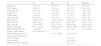
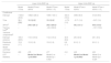
ResultsParticipants' characteristics are presented in Table 1. Effects of moment, maturation, and training load on BMC of the limbs are presented in Table 2. A significant effect of time was observed for BMC of the lower limbs. From an estimated 1183.8 g (SE = 40.6 g) of initial leg BMC, a significant rate of change of 12.1 g/week (SE = 2.6 g/week) across the three moments was observed. Adolescents at post-PHV had a significant 245.6 g (SE = 56.1 g) higher BMC at legs compared to adolescents at circa-PHV. Maturation did not alter the rate of bone accrual during the period investigated. No effects of internal training load were observed.
Sample characteristics over the training period (n = 17).
YPHV: years from peak height velocity. BMI: body mass index. BMC: area bone mineral content. aBMD: area bone mineral density.
MVPA, Moderate-to-Vigorous physical activity.
Effects of time, maturation, and training load on bone mineral content of lower and upper limbs (n = 17).
| Lower limbs BMC (g) | Upper limbs BMC (g) | |||||
|---|---|---|---|---|---|---|
| Model 1Time | Model 2Time + APHV | Model 3Time + APHV + ITL | Model 1Time | Model 2Time + APHV | Model 3Time + APHV + ITL | |
| Coefficients | ||||||
| Intercept | 1183.8 (40.6) | 1082.6 (36.3) | 1094.7 (40.1) | 333.3 (10.0) | 308.9 (8.9) | 312.9 (9.6) |
| Time | 12.1 (2.6)⁎ | 12.2 (2.6)⁎ | 12.2 (2.6)⁎ | −0.79 (1.6) | −0.71 (1.6) | −0.67 (1.6) |
| APHV# | – | 245.6 (56.1)⁎ | 262.4 (60.6)⁎ | – | 59.2 (13.1)⁎ | 64.8 (13.8)⁎ |
| Training load## | – | – | −40.4 (59.8) | – | – | −13.7 (13.6) |
| Variance components$ | ||||||
| Intercept variance | 165.9 (28.5) | 113.5 (19.6) | 112.0 (19.3) | 38.9 (6.8) | 25.9 (4.6) | 25.1 (4.5) |
| Residuals variance | 13.9 (1.7) | 13.9 (1.8) | 13.8 (1.8) | 8.9 (1.1) | 8.9 (1.1) | 8.9 (1.1) |
| Model summary | ||||||
| Deviance | 490 | 478 | 477 | 414 | 400 | 400 |
| LRT Statistics | – | Model 2vs Model 1p=0.0003⁎ | Model 3 vs Model 2p = 0.502 | – | Model 2vs Model 1p=0.0002⁎ | Model 3 vs Model 2p = 0.324 |
Values are estimates (standard error). BMC, bone mineral content. APHV, age from peak height velocity. ITL, internal training load. LRT, likelihood-ratio test.
No significant effect of time was observed for BMC of the upper limbs. From an estimated 333.3 g (SE = 10.0 g) no significant rate of change was observed across the three moments investigated. Adolescents at post-PHV had a significant 59.2 g (SE = 13.1 g) higher BMC at the arms compared to adolescents at circa-PHV. Maturation did not alter the rate of bone accrual during the period investigated. No effects of internal training load were observed.
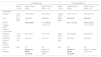
The effects of time, maturation, and training load on the BMC of the trunk and body are presented in Table 3. A significant effect of time was observed for BMC of the trunk. From an estimated 739.1 g (SE = 27.9 g) a significant rate of change of 12.1 g/week (SE = 3.1 g/week) in BMC was observed. Adolescents at post-PHV had a significant 158.5 g (SE = 39.9 g) higher BMC at the trunk compared to adolescents at circa-PHV. Maturation did not alter the rate of bone accrual during the period investigated. No effects of internal training load were observed. Similarly, a significant effect of time was observed for BMC of the body. From an estimated 2682.8 g (SE = 78.2 g) a significant rate of change of 26.4 g/week (SE = 3.8 g/week) was observed. Adolescents at post-PHV had a significant 451.1 g (SE = 114.3 g) higher BMC at the trunk compared to adolescents at circa-PHV. Maturation did not alter the rate of bone accrual during the period investigated. No effects of internal training load were observed.
Effects of time, maturation, and training load on bone mineral content of trunk and total body (n = 17).
| Trunk BMC (g) | Total body BMC (g) | |||||
|---|---|---|---|---|---|---|
| Model 1Time | Model 2Time + APHV | Model 3Time + APHV + ITL | Model 1Time | Model 2Time + APHV | Model 3Time + APHV + ITL | |
| Coefficients | ||||||
| Intercept | 739.1 (27.9) | 673.7 (26.3) | 687.2 (28.3) | 2682.8 (78.2) | 2497.0 (73.7) | 2521.5 (81.4) |
| Time | 12.1 (3.1)⁎ | 12.1 (3.1)⁎ | 12.2 (3.1)⁎ | 26.4 (3.8)⁎ | 26.5 (3.8)⁎ | 26.5 (3.4)⁎ |
| APHV# | – | 158.5 (39.9)⁎ | 177.3 (42.3)⁎ | – | 451.1 (114.3)⁎ | 485.0 (123.6)⁎ |
| Training load## | – | – | −45.3 (41.7) | – | – | −81.7 (121.9) |
| Variance components$ | ||||||
| Intercept variance | 112.0 (19.4) | 80.3 (14.0) | 77.6 (13.6) | 320.8 (55.1) | 231.6 (39.8) | 228.5 (39.3) |
| Residuals variance | 16.6 (2.1) | 16.5 (2.1) | 16.6 (2.1) | 20.8 (2.6) | 20.8 (2.6) | 20.9 (2.6) |
| Model summary | ||||||
| Deviance | 488 | 477 | 476 | 538 | 527 | 527 |
| LRT Statistics | – | Model 2vs Model 1p=0.008⁎ | Model 3 vs Model 2p = 0.286 | – | Model 2vs Model 1p=0.009⁎ | Model 3 vs Model 2p = 0.505 |
Values are estimates (standard error). BMC, area bone mineral content. APHV, age from peak height velocity; ITL, internal training load; LRT, likelihood-ratio test.
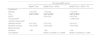
The effects of time, maturation, and training load on total body BMD are presented in Table 4. A significant effect of time was observed for total body BMD. From an estimated 1.16 g/cm2 (SE = 0.02 g/cm2) a significant rate of change of 0.007 g/cm2/week (SE = 0.003 g/cm2/week) was observed. No significant effects of aPHV and training loads were observed for BMD.
Effects of time, maturation, and training load on total body bone mineral density (n = 17).
| Total body aBMD (g/cm2) | |||
|---|---|---|---|
| Model 1Time | Model 2Time + APHV | Model 3Time + APHV + ITL | |
| Coefficients | |||
| Intercept | 1.18 (0.02) | 1.16 (0.02) | 1.16 (0.02) |
| Time | 0.007 (0.003)⁎ | 0.007 (0.003)⁎ | 0.007 (0.003)⁎ |
| APHV# | – | 0.06 (0.03) | 0.06 (0.04) |
| Training load## | – | – | −0.009 (0.04) |
| Variance components$ | |||
| Intercept variance | 0.07 (0.01) | 0.07 (0.01) | 0.06 (0.01) |
| Residuals variance | 0.02 (0.002) | 0.02 (0.002) | 0.02 (0.002) |
| Model summary | |||
| Deviance | −185 | −188 | −188 |
| LRT Statistics | – | Model 2 vs Model 1p = 0.0856 | Model 3 vs Model 2p = 0.806 |
Values are estimates (standard error). aBMD, area bone mineral density. APHV, age from peak height velocity; ITL, internal training load; LRT, likelihood-ratio test.
The purpose of this study was to analyze aBMD and BMC accrual at different skeleton sites in male adolescent footballers across a short 15 weeks of training and competition. The main findings were: 1) 15 weeks of football increased BMC of the lower, but not of the upper skeleton; 2) Maturation has a significant impact on BMC, as post-pubertal players had higher BMC than the pre-pubertal counterparts; 3) no effects of internal training load on bone outcomes were observed. The present investigation adds novel data to the literature indicating that even a short 15-week period of football can increase bone mass in the legs and trunk with no effect on the accumulated perceived training load in adolescents participating in their first competitive season.
Regarding adolescents' bone mass in the present study, at baseline participants had higher total body BMC and aBMD (BMC = 2711.1 g and aBMD = 1.18 g/cm2) compared with reference values for 14–16 years old adolescents’ non-athletes at the 90th percentile of previously published work (BMC = 2350.0 g and aBMD = 1.15 g/cm2).24 These values may reflect athletes' prior sport involvement through school training and competition. Despite presenting an elevated bone mass compared to reference values, participants also gained significant amounts of bone across the investigated 15 weeks. These results are in line with findings available in the literature on football participation with longer follow-ups and DXA-assessed bone variables. For example, Agostinete et al.,18 demonstrated that after 9 months of follow-up, adolescent footballers enhanced aBMD at different body sites. In addition, the gains in BMD and BMC among football practitioners were higher compared to adolescents of the non-sports control group,1,13 and the non-impact sports practiced by swimmers and cyclists.15,25
The positive impact of football on bone tissue in the lower limbs and trunk but not in the arms can be explained by the characteristics of the sport, such as running, accelerating, braking, jumping, changing direction, passing, and kicking.25 These actions generate direct stimulus on the bone mechanoreceptors in the legs, but not in the arms.26 Moreover, football engagement requires high quadriceps and hamstring muscle activity, generating an indirect stimulation of the bone tissue.25 A recent systematic review highlighted that the lower limbs suffer the highest impacts of football during growth.2 In addition, it is likely that beginning to train and compete in a professional U-15 team contributed to increases in bone mass due to the systematic training occurring at this level, as observed in the present study. In fact, similarly, previous work has evidenced a high increase in bone mass at the beginning compared to later years of training in pre-pubertal youth.27
A positive maturation effect was observed for BMC, BMD, and muscle mass in accordance with the literature.5,28 This is because, in the stages of maturational development, peak height velocity precedes the peak of lean mass and the peak of bone mass accrual, while the peak of lean mass also precedes the peak of bone mass accrual.3,29 Muscle mass is a strong determinant of bone mass 12 due the strains generated by muscle contractions stimulating the skeleton to adaptations.27,30 Therefore, post-PHV is the period of high bone mass accrual, and these results highlight the importance of stimulating the bone tissue in circumpubertal years. However, no interaction between maturation and training weeks was observed indicating that the rate of bone mass accrual was similar between the groups of maturation, with football influencing equally the skeleton of boys at circa and post-PHV.
While other studies have reported an inverse association between training loads and bone mass accrual in swimmers,18 in the present investigation no effects of the accumulated training load were observed in the different skeleton sites. Different from the present study, swimmers with a high training load experienced a lower bone mass accrual due to the characteristics of swimming. In fact, a study has demonstrated that swimmers have lower BMC compared to sedentary controls,12 and a higher swimming training load can be detrimental to bone accrual during adolescence. However, studies analyzing the effects of different training variables in football are scarce the impact of training load is unknown.
Among the few studies available in the literature with a similar follow-up to the present study, increased bone density of trabecular and cortical compartments following an increased volume of training over 12 weeks in elite athletes was demonstrated.17 Thus, it was hypothesized that bone adaptation to training prevents structural fatigue and possible stress fractures. However, no studies have yet tested this hypothesis using measures of training load, impeding comparisons with the current findings. Therefore, when considering that both training intensity and volume seem to affect bone outcomes,26 it is likely that other measures of training intensity, including jumps, acceleration and PlayerLoad can better capture the impact of football training on bone outcomes. Future studies are encouraged to consider the short-term effects of football on different bone outcomes and the effect of different measures of training load.
The present study has some limitations. Firstly, the control group was not included precluding comparisons between groups. Secondly, despite being athletes' first competitive season, their prior contact with school-based sports could potentially have introduced uncontrolled residual effects. Moreover, calcium intake was obtained at one-time point. However, despite a low calcium intake, participants had bone mass > 90th percentile for their age.30 Future studies measuring right and left sites may contribute to the understanding of bone mass acquisition between dominant and non-dominant limbs.
A few strengths are worth mentioning. The inclusion of a short follow-up in a competitive season, which had not been explored in the literature. Also, participants were starting their professional football careers, allowing us to investigate how initiation to football can alter the rate of bone gains. Altogether, the present findings add novel data to the literature showing that shorter periods of competitive football impact bone outcomes of adolescents, as observed in studies with longer follow-ups.
A short 15 weeks of football training and competition caused a significant increase in BMC in the legs and trunk of adolescent footballers who started to train and compete. Adolescents post-PHV had higher BMC and aBMD, but maturation did not change the rate of gain in bone mass. Finally, no effects of training load were observed.


