
Inborn Errors of Immunity are characterized by infectious conditions and manifestations of immune dysregulation. The diversity of clinical phenotypes can make it difficult to direct the laboratory investigation. This article aims to update the investigation of immunological competence in the context of primary defects of the immune system.
Source of dataSearches were carried out on Pubmed to review articles published in the last five years, in English, French or Spanish, using the terms “diagnosis” OR “investigation” AND “immunodeficiency” or “primary immunodeficiency” or “inborn errors of immunity” NOT “HIV”. Recent textbook editions have also been consulted.
Summary of findingsThe immune system competence investigation should be started based on clinical phenotypes. Relevant data are: characterization of infectious conditions (location, recurrence, types of infectious agents, response to treatment), age during symptom onset and associated manifestations (growth impairment, allergy, autoimmunity, malignancies, fever and signs of inflammation without the identification of infection or autoimmunity) and family history. These data contribute to the selection of tests to be performed.
ConclusionsThe diagnostic investigation of Inborn Errors of Immunity should be guided by the clinical characterization of patients, aiming to optimize the use of complementary tests. Many diagnoses are attained only through genetic tests, which are not always available. However, the absence of a diagnosis of certainty should never delay the implementation of therapeutic measures that preserve patient life and health.
Primary immune system defects are traditionally called primary immunodeficiencies (PID). However, due to the large number of defects recently described, which include manifestations that are more related to the immune system dysregulation than to its deficiency, this nomenclature has been replaced by Inborn Errors of Immunity (IEI).1,2
The current classification consists of 416 diseases caused by mutations in more than 450 genes.2 The infectious manifestations are the most common ones. In recent years, primary defects of the immune system have been associated not only to frequent or severe infections, but also to the specific susceptibility to certain infectious agents. Moreover, there has been a better identification of other clinical features, such as severe allergies, inflammatory processes, lymphoproliferation, autoimmunity and even malignancies.3
Due to the diversity of clinical manifestations, it is important that the performance of the laboratory investigation of IEI be directed based on the clinical phenotype and an initial immunological screening.4 Several studies have shown the importance of family history as a warning sign. Complementary tests should follow a sequence of increasing complexity, starting with a simple blood count and measurement of immunoglobulin levels, to the complete sequencing of the exome or genome, using the new generation sequencing technique.5,6
Unfortunately, many of the tests required to elucidate the diagnosis of diseases in this group are not available in commercial laboratories, some not even available in research laboratories in our country. In regards to the existing tests in commercial laboratories, we still have the problem of them not being included in the list of tests covered by the Brazilian Unified Health System (SUS) or Supplementary Health system.
ObjectivesOur objective was to develop a proposal for an updated approach to the laboratory investigation of immunological competence in the context of IEI.
Source of dataWe carried out searches on Pubmed to review articles published in the last five years, in English, French or Spanish, using the terms “diagnosis” OR “investigation” AND “immunodeficiency” or “primary immunodeficiency” or “inborn errors of immunity” NOT “HIV”. Recent textbook editions have also been consulted.
ResultsInitial assessmentIt is important to note that the approach described below will focus on the immunological tests, although other laboratory and imaging tests are relevant for the investigation of the non-infectious manifestations of these diseases.
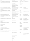
The characterization of infectious conditions (location, recurrence, types of infectious agents, response to treatment, adverse reactions to vaccines), age during symptom onset and associated manifestations (growth impairment, allergy, autoimmunity, lymphoproliferation, malignant diseases, fever and signs of inflammation without identification of infection or autoimmunity), allow directing the immune response sector most likely to be affected, which should be primarily evaluated.7Table 1 shows the initial complementary tests according to the type of infection, the identified infectious agents and associated non-infectious manifestations.
Immunological evaluation according to the type of infection, infectious agent and associated manifestations.
| Infections | Other manifestations | Suspected IEI | Initial tests |
|---|---|---|---|
| Severe, disseminated by viruses, bacteria, fungi, protozoa, opportunistic microorganismsCytomegalovirus,Herpesvirus,MycobacteriaCandida sp,Aspergillus sp,ToxoplasmosisP. jirovecii,Cryptosporidium spReaction to BCG vaccine | Early onsetGrowth impairmentChronic diarrheaAbsence of thymusMicrocephaly, with or without facial dysmorphism | Combined T and B cell immunodeficiencies - SCID | CBC, lymphocyte count (CD3, CD4, CD8, CD19, CD56),TREC, immunoglobulin measurement |
| Facial malformations and dysmorphismsBone dysplasiaFracturesScoliosisShort statureMicrocephaly Bruising / bleedingCerebellar ataxiaOculocutaneous TelangiectasiasGlobal development delayIntellectual deficitJoint hypermotilityExtensive eczemaIchthyosisEctodermal dysplasia | Combined immunodeficiencies associated with syndromes | ||
| Severe and / or recurrent bacterial infections, encapsulated respiratory, articular microorganisms, sepsis, Pneumococcus,Hemophilus,MycoplasmaGiardia lambliaIntestinal bacteria: Campylobacter sp, Salmonella spEnterovirus (polio vaccine)Norovirus | Symptom onset above 6 monthsAbsence of tonsils / adenoidsLymphoproliferationLymphoid nodular hyperplasiaDevelopmental delayChromosomal disorders | Predominantly antibody defects | Complete blood count,Immunoglobulin measurements,B lymphocyte count,KREC |
| Bacterial, encapsulated microorganisms of the respiratory tract, articular or meninges, sepsisPneumococcus, Neisseria sp, Hemophilus | Manifestations of autoimmunity, lupus-like,glomerulopathy | Complement defects | C3, C4, CH50 |
| Non-significant, non-severeCandidiasisBacterial infectionsEpstein-Barr virus | Early onsetLymphoproliferationHemophagocytic lymphohistiocytosisPartial oculocutaneous albinismDevelopmental delayAutoimmune cytopeniasAutoimmune endocrinopathiesSevere inflammatory bowel disease | Diseases with immune dysregulation | Blood countHematoscopyAutoantibodies |
| Recurrent skin infectionsDeep abscessesNecrotizing pneumoniaOsteomyelitisCatalase positive microorganisms:Staphylococcus sp, Klebsiella sp, Serratia sp, Pseudomonas sp,Aspergillus spCandida sp,Burkholderia cepacia,M. tuberculosis | Early onsetGranulomasNonspecific inflammatory bowel diseasePancreatic insufficiencyDevelopmental delayIntellectual deficitMouth ulcersDelayed umbilical cord fallPoor healingPeriodontal diseasePulmonary alveolar proteinosisLymphedemaMyelodysplasia | Numeric or functional phagocyte diseases | Blood countMorphological evaluation of neutrophils in HematoscopyNeutrophil oxidative burst test (DHR) |
| Recurrent infections by only one type or few types of microorganism | Little or no fever | Innate immunity deficiency –IRAK4/MyD88 defect | CBC, immunoglobulin measurement, lymphocyte phenotyping, CH50 to rule out other diagnoses, |
| Bacterial, invasive, sepsis, | Delayed umbilical cord fall | CD62L Shedding assay | |
| meningitis, arthritis, | Improves with age | ||
| osteomyelitis | |||
| Pneumococcus | Absence of spleen | ||
| Recurrent infections by only one type or few types of microorganisms | Reaction to BCG vaccine | Mendelian susceptibility to mycobacteria | Functional assessment of the IFN γ-IL12 axis |
| Mycobacteria | |||
| Intracellular fungi and bacteria | |||
| Recurrent infections by only one type or few types of microorganisms | No other manifestations | Innate immunity deficiency with susceptibility to viral infections | Immunoglobulin measurement |
| HPV-disseminated warts with or without bacterial infections | Blood count | ||
| Recurrent herpes simplex encephalitis | Lymphocyte phenotyping to rule out other diagnoses, | ||
| Severe reactions to triple viral and yellow fever vaccines | CD62L Shedding assay | ||
| Recurrent infections by only one type or few types of microorganisms | Ectodermal dysplasia | Innate immunity deficiencies with susceptibility to fungi | CBC, immunoglobulin measurements, lymphocyte phenotyping, DHR to rule out other diagnoses |
| Cutaneous and / or invasive fungal infections, mainly Candida sp, with or without cutaneous staphylococcus infections | Autoimmune endocrinopathies | ||
| Non-significant, non-severe, nor recurring | Inflammatory manifestations in the absence of infectious agents or autoimmunity | Autoinflammatory diseases | Inflammatory activity test |
| Recurrent fever | Autoantibodies, immunoglobulin measurements and lymphocyte phenotyping to rule out other diagnoses | ||
| Urticarial rash without pruritus | |||
| Panniculitis | |||
| Lipodystrophy | |||
| Lytic bone lesions | |||
| Arthritis | |||
| Pyoderma gangrenosum | |||
| Severe acne | |||
| Pustular psoriasis | |||
| Encephalopathy | |||
| Strokes | |||
| Pernio |
It is important to keep in mind that a crucial initial step is to rule out the presence of any secondary immunodeficiency, particularly HIV infection, and also those caused by the use of drugs or renal and gastrointestinal losses.7
A simple blood count with hematoscopy provides much information that may be relevant to guide the diagnostic investigation: presence of anemia, thrombocytopenia, with or without small platelets, leukocytosis >20,000, lymphopenia, neutropenia, monocytopenia, eosinophilia and/or giant granules in neutrophils.7 Lymphopenia, defined by a lymphocyte count <2500 in the first months of life, may indicate impaired cell immunity.5
The measurement of IgG subclasses is a test of questionable relevance and may be important for a patient with IgA deficiency who has significant recurring bacterial infections, but it should always be associated with the assessment of the functional antibody response.5,7
The measurement of isohemagglutinins in children over one year of age and who do not belong to the AB blood group can help in assessing the ability to produce antibodies, as they are natural IgM antibodies against the polysaccharide antigens blood group.7 To assess the response to protein antigens, the vaccine response is evaluated and then we usually request serological tests for measles, rubella, mumps, tetanus, poliovirus and diphtheria. In patients who are already receiving immunoglobulin replacement, the option in our country would be to assess the response to the rabies vaccine.5 The response to polysaccharide antigens should be obtained by requesting IgG measurements for 23 pneumococcal serotypes before and 4–6 weeks after the application of the unconjugated 23-valent pneumococcal vaccine. A normal response to polysaccharides is considered to be the presence of titers > 1.33 in the post-vaccine measurement, or a two-fold increase in relation to pre-vaccine titers, for more than 50% of the eleven serotypes in children under six years of age and 70% in those over the age of six (2, 8, 9N, 10A, 11A, 12F, 15B, 17F, 20, 22F and 33F). It is important to use the serotypes absent from any of the conjugate vaccines applied in the first year of life.7–9 The evaluation of only seven serotypes present in any of the conjugate vaccines is not appropriate for this diagnosis.
The measurement of serum immunoglobulins is a screening test not only for antibody-mediated immunity, but also for some combined defects of B (CD19+ or CD20+) and T cells (CD3+), especially if we associate it with the evaluation of a vaccine response. Immunodeficiencies associated with syndromic conditions can also be screened through this evaluation, such as in hyper IgE syndrome, ataxia-telangiectasia or ectodermal dysplasia.5
Tests that are widely used for the initial immunological evaluation, as immunoglobulin A, M, G and E measurements, and the lymphocyte subpopulation counts (CD3, CD4, CD8, CD19, CD56, respectively T, T4, T8, B and NK) should be compared with reference values for age and, preferably, in the same population to which the patient belongs.5
Addressing specifically the T-lymphocyte-mediated immunity, intradermal hypersensitivity tests can be used to screen for cell immunity. The antigens used are: candidin, streptokinase/dornase, staphylococcal toxin, trichophytin and PPD. Patients older than one year must respond (3-mm papule) to at least one of the tested antigens in 48–72 h. This type of assessment has been abandoned, as access to flow cytometry became easier when the monitoring of HIV-positive patients was implemented. The functional assessment of T lymphocyte-mediated immunity can be performed in vitro. Stimulation can be performed with mitogens and antigens, traditionally by incorporating tritiated thymidine or, more recently, by incorporating a nucleotide (EdU) via the fluorescence system or using dye (CSFE).5,7 This test has restricted availability. When unavailable, intradermal in vivo tests can be useful.
The evaluation of TREC (T-cell Receptor Excision Circles) and KREC (Kappa-deleting Recombination Excision Circles), the counting by PCR of excision circles that are discarded during the process of surface receptor rearrangement of T and B cells respectively, constitute an interesting screening test performed using the collection of Dried Blood Spots(DBS)on filter paper.10 TREC allows assessing the presence of naïve T cells that recently left the thymus and KREC assesses the presence of immature B lymphocytes. Generally used in the neonatal screening for T and B cell defects, this count can also be useful in the investigation of an IEI. 11 When a Severe Combined Immunodeficiency is suspected, additional labeling of NK cells (CD16 + CD56 +) makes it possible to assume the associated molecular defects.4
Considering neutrophil defects, congenital neutropenia can be assessed, as described, by a leukogram. However, cyclic neutropenia requires weekly serial leukograms (two to three times a week for six weeks) to detect periods of marked descrease of neutrophils counts.12,13 The functional assessment should be requested according to the clinical history, and the NBT (nitrobluetetrazolium) or DHR (Dihydrorodamine) test can be applied for intracellular death defects such as Chronic Granulomatous Disease. Other stages of phagocyte activation, the neutrophil chemotaxis, can be assessed using the Boyden chamber, with the reading being performed using a common microscope or on an agar medium; it is, however, a laborious technique with a difficult standardization, performed in research centers.5,14
The activation of the classical and alternative complement pathways can be assessed using CH50 or CH100 and through AP50. These tests allow targeting the possibly deficient components in these pathways. Although the measurement of components C3 and C4 are readily available, complete primary deficiencies of these proteins are rare. For the diagnosis of lectin pathway deficiencies such as MBL (mannose-binding lectin) or MASP2 (MBL-associated serine protease-2), it is necessary to carry out specific assays for this purpose. It is important to emphasize that altered values when evaluating the complement system should be verified, since they are acute phase and extremely thermolabile proteins, and the consumption in the presence of an acute condition or sample collection/handling problems are common.7,15
Flow cytometry evaluationFlow cytometry has revolutionized immunology and the study of IEI.16 As mentioned before, the use of this device was driven by the AIDS epidemic and it has allowed the detection of the expression of surface, intracellular or intranuclear proteins, which allows the identification of cell populations and subpopulations, the detection of biological effects of cell activation or those due to immunological defects, as well as the assessment of the immune system function.17 Therefore, in addition to cell quantification, many functional assays can be performed, providing rapid results.16,17
The most commonly requested evaluation is basal lymphocyte phenotyping: B (CD19 or CD20), NK (CD56/16), T (CD3), T4 (CD3CD4) and T8 (CD3CD8). However, several situations below can demonstrate the scope of this method. In cases of hypogammaglobulinemia with a normal number of B lymphocytes (CD19+), it is important to evaluate sub-populations of B lymphocytes: naïve (CD27-), memory (CD27+), with class switch recombination (IgM-) or without class switch (IgM +).5,17
In combined T and B cells defects without intense lymphopenia, it is necessary to evaluate more sub-populations of T lymphocytes: naïve (CD3+ CD45RA+), memory (CD3+ CD45RO+), double negative CD3 + TCRαβ CD4-CD8-) and Th17.5,17
The labeling of subpopulations of NK cells (CD56 and CD16) permits the identification of five populations: CD56 bright CD16 -, CD56 bright CD16dim, CD56 dim CD16-, CD56dim CD16+ and CD56-CD16 + . CD56 bright NK cells are the ones with highest production of IFNγ, whereas CD56 dim cells have the highest amounts of perforin and granzymes, while CD16+ cells act in antibody-mediated cytotoxicity.5,17 NK T cells have CD3 and CD56, act on innate immunity and are altered in lymphoproliferative syndromes associated with the Epstein-Barr virus.
In fact, the cell subpopulations identified by flow cytometry are much more numerous than those shown herein and have allowed not only a better understanding of the IEI, but also the immune system function.16,18
It is possible to label, also by flow cytometry, the expression of proteins such as BTK, WASp, CD40/CD40L, CD11/CD18 and IL-10R, assisting in the diagnosis, respectively, of X-linked agammaglobulinemia, Wiskott-Aldrich syndrome, Hyper IgM syndrome, leukocyte adhesion defect type I, and early and severe inflammatory bowel disease due to IL-10R defect. A large number of other molecules can also be labeled.17,18
Additionally, functional in vitro assays can be performed by flow cytometry to assess lymphocyte proliferation and activation, TCD8 and NK cytotoxicity, IFN-γ-IL-12 axis activity, oxidative burst in phagocytes (Dihydrorodamine, DHR) and specific activation of TLR and many others.17,18
Advanced assessmentThe tests used for the initial evaluation allow the identification of a considerable percentage of patients with IEI. However, they do not allow the diagnosis of many other defects, which have been more recently described. The performance of further specialized tests, which, in general, are not part of the laboratory routines, may be necessary.
The evaluation of the repertoire of T cell receptors (TCR) evaluating the β chain of the variable region (Vβ) can be performed by flow cytometry or by PCR, identifying its distribution curve, oligo (pathological) or polyclonal (normal).5 Thus, we identify diseases in which the number of lymphocytes is normal but with a cell response that is impaired due to the restriction of its specificity and its ability to recognize different antigens.
The measurement of the enzymatic activity of adenosine deaminase (ADA) and purine nucleoside phosphorylase (PNP) in peripheral blood can assist in the diagnosis of cases of severe combined immunodeficiency (SCID); these defects, in turn, can be treated with enzymatic replacement until the definitive treatment can be carried out with hematopoietic stem cell transplantation. An easy-to-perform test is the measurement of uric acid, which is extremely low in PNP deficiency and acts on purine metabolism. The alpha-fetoprotein level is high in patients with ataxia-telangiectasia, which is a good test to be used when the disease is suspected.7
Cytotoxicity assays are useful when defects in NK cells are suspected with major viral infections, particularly by the human papillomavirus (HPV) or the herpesvirus family. They can be performed by assessing the release of chromium51 in the supernatant from the lysed target cell or the expression of CD107a on the cell surface. In fact, the first method assesses the cytotoxicity itself, and the second one evaluates the degranulation capacity. In perforin defects, for instance, there is normal expression of CD107a on the NK surface, but little elimination of chromium51 in the supernatant.5
Functional assays for the evaluation of defects in the IFNγ-IL-12 axis, which cause Mendelian susceptibility to mycobacteria, are performed by evaluating IL-12 production in vitro, after stimulating the peripheral mononuclear cells with IFNγ, as well as assessing the IFNγ production after lymphocyte stimulation with IL-12.5
The functional assessment of Toll Like Receptors (TLR), relevant in the case of susceptibility restricted to one type of infectious agent suggesting an inate immunity deficiency, is carried out using the CD62L shedding assay. Using several specific stimuli for different TLR, the amount of CD62L released by neutrophils in the supernatant is evaluated. The increase in CD62L identifies healthy signaling pathways.5 Serum type I IFN measurement is useful when investigating a group of autoinflammatory diseases, called interferonopathies.7,19
Genetic testingChromosomal microarray analysis is a technique widely used in genetics, particularly in the presence of a syndromic or unspecific phenotype, aiming to identify one or a few suspect genes.20 This test identifies chromosomal losses and gains, copy number variants (CNV) throughout the genome, deletions and duplications with greater sensitivity than the karyotype. It also has the capacity to detect excess homozygosity, suggesting consanguinity that is sometimes restricted to a certain part of the genetic code, which helps in directing the search for mutations by new generation sequencing. The method is widely used to identify the 22q11 deletion, related to DiGeorge Syndrome (thymic aplasia or hypoplasia, congenital heart disease, associated hypoparathyroidism).21
Genetic sequencing using the Sanger method is still considered the gold standard for the diagnosis of mutations.20,21 However, the new generation sequencing (NGS), adopted since 2010, allows the simultaneous investigation of many more genes and at a much lower cost. The NGS is carried out using gene panels, chosen according to the type of disease to be investigated, through the Whole Exome Sequencing (WES), in which only the part of the genome that encodes proteins (exons) is sequenced, or using the Whole Genome Sequencing (WGS). The WGS has a greater diagnostic capacity; however, due to the high cost and the difficulty in handling a great deal of information, it is not yet used in clinical practice. Sequencing using gene panels is more affordable; however, it may not allow diagnosis in many cases. The WES is an intermediate option in terms of cost and amount of data to be analyzed and has been considered the most cost-effective test.22 Regardless of the type of sequencing to be performed, it is essential that a good clinical and immunological characterization of the patient be performed, in addition to a complete family history, aiming to define the main suspect genes and possible forms of inheritance.23
Genetic sequencing has allowed many diagnoses to be established, describing new defects and even enabling a better knowledge of the immune system function. Genetic tests have provided the definitive diagnosis of most IEIs. However, it has also brought many challenges regarding cost, access and interpretation, the need to improve the analysis of the obtained data and to prove the pathogenicity of some mutations of uncertain significance to this date.21,22 In the latter case, functional tests, which are often performed using cytometry, are necessary to prove the pathogenicity.18
ConclusionsA standard listing of immunological tests should be avoided. It is necessary to optimize laboratory test requests based on the clinical characterization of patients.
There are many advanced tests of difficult access or that are unavailable in our country. Many diagnoses in the IEI group are only possible with genetic testing, which is not always available. Despite the advantages of having an accurate diagnosis for patient management, its absence should never delay the implementation of therapeutic measures that maintain the patients' life and health until their diagnosis is confirmed.
Conflicts of interestThe authors declare no conflicts of interest.


